Review
. 2008 Jul;9(6):471-86.
doi: 10.2174/138920008784892065.

Structure/function evaluations of single nucleotide polymorphisms in human N-acetyltransferase 2

Affiliations
Review

Structure/function evaluations of single nucleotide polymorphisms in human N-acetyltransferase 2

Jason M Walraven et al. Curr Drug Metab. 2008 Jul.

Abstract

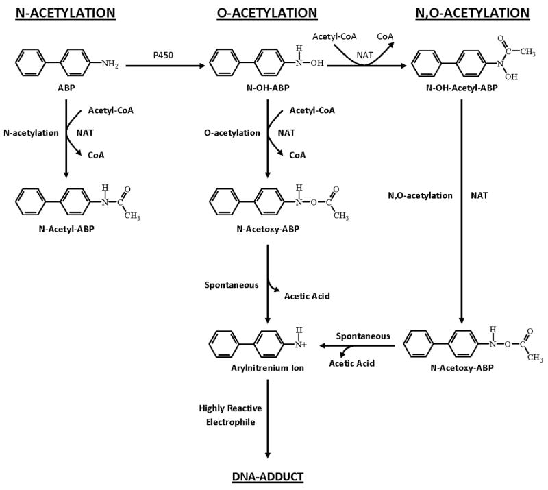
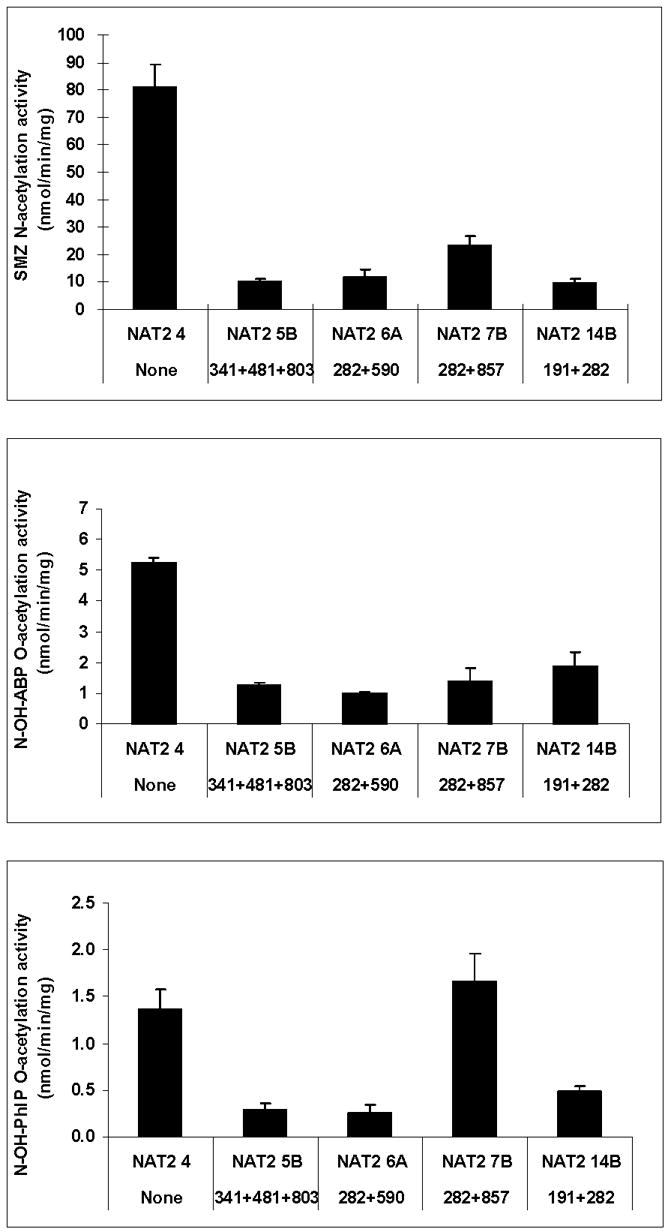
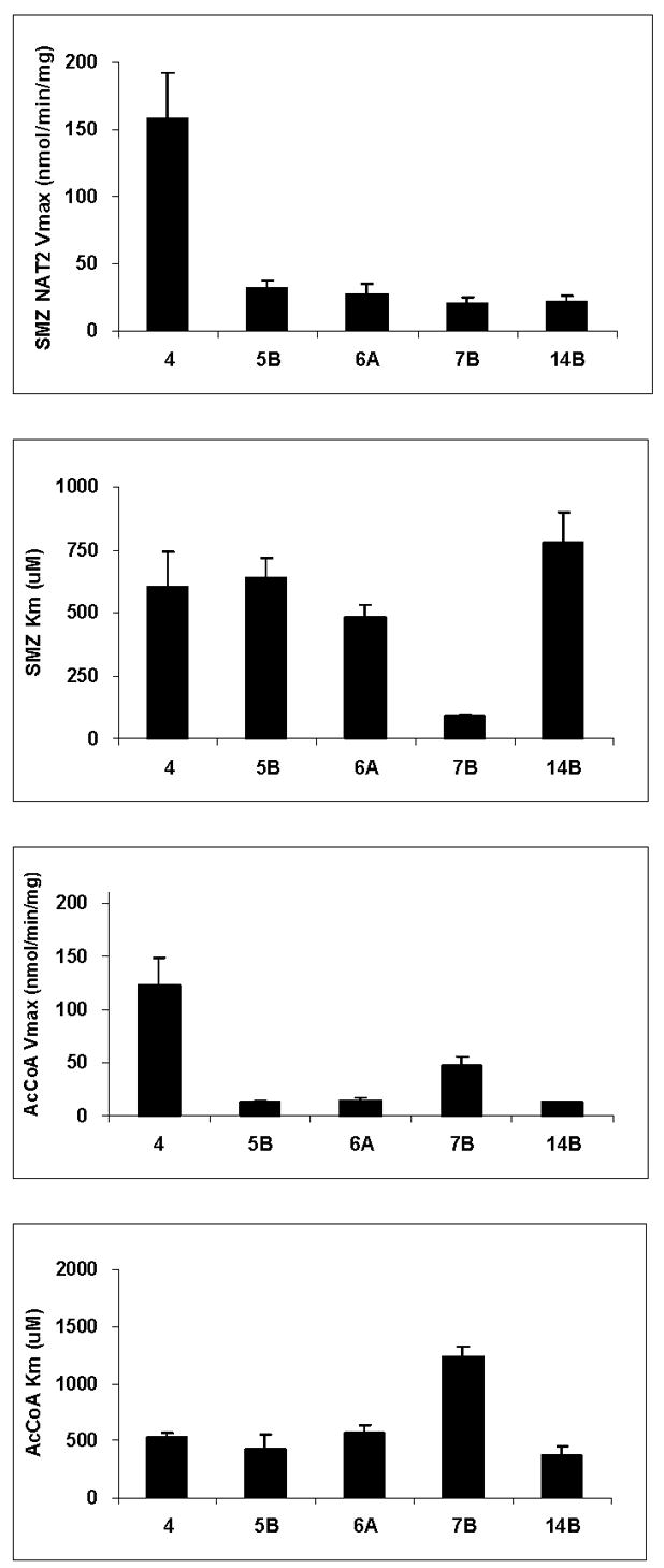
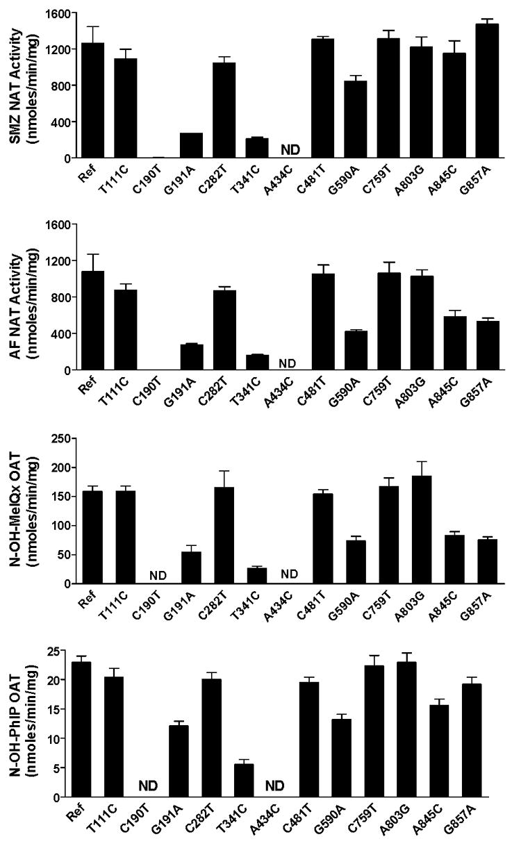
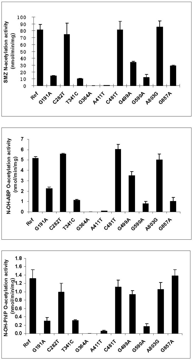
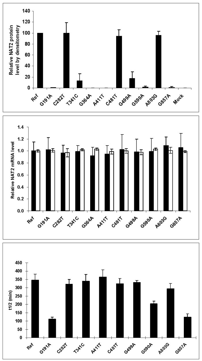
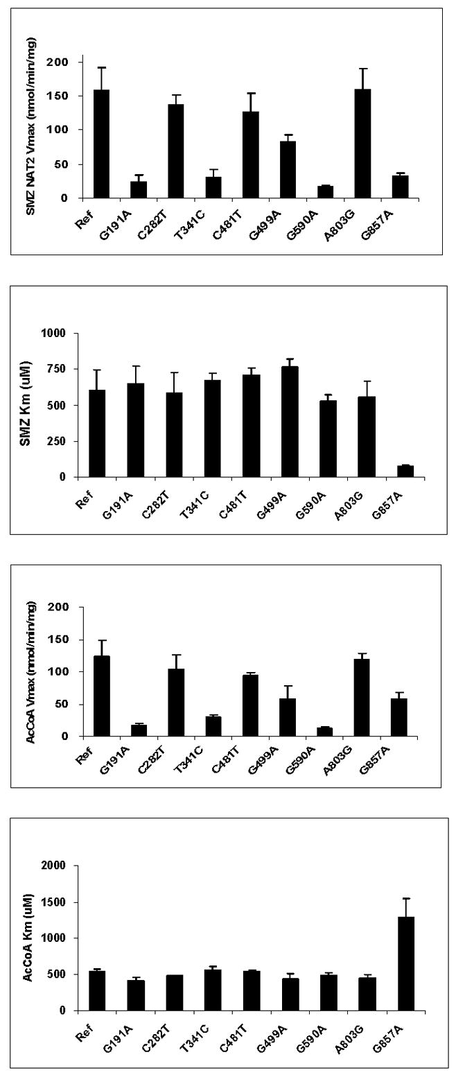
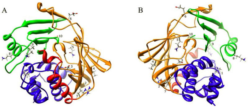
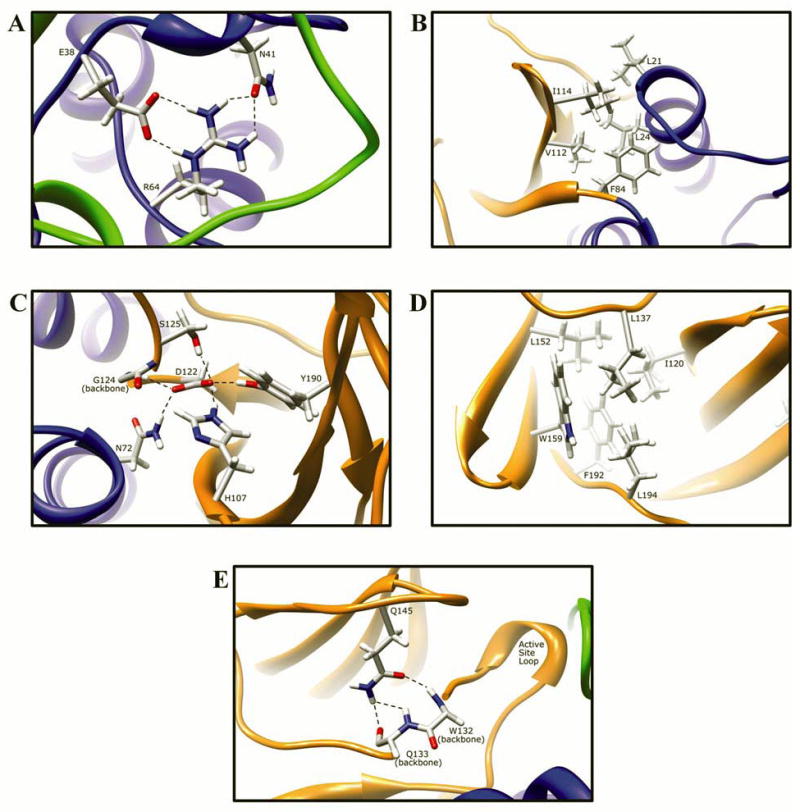
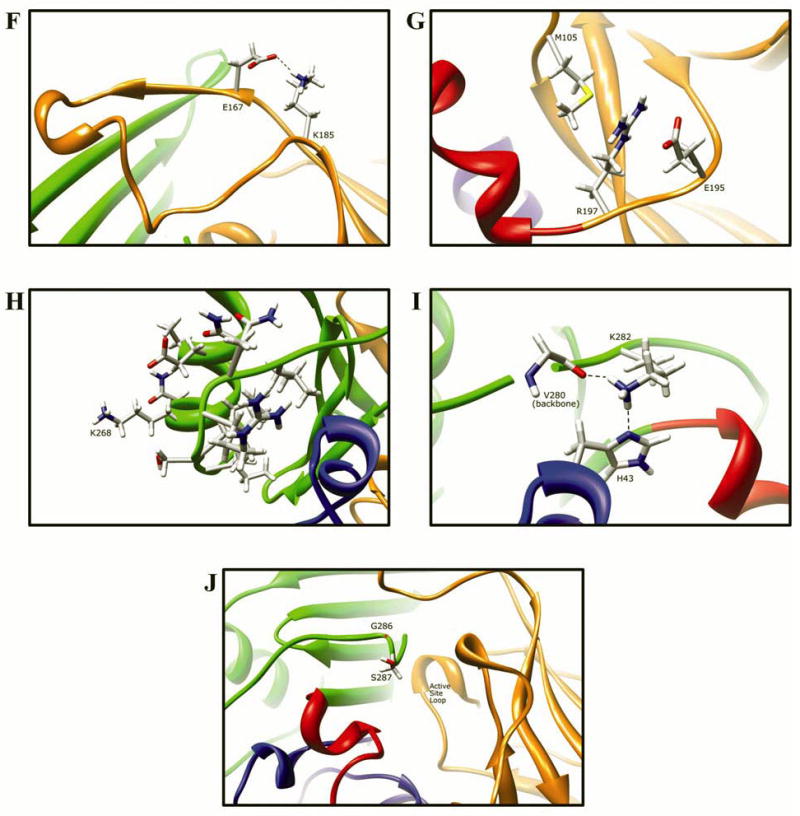
Arylamine N-acetyltransferase 2 (NAT2) modifies drug efficacy/toxicity and cancer risk due to its role in bioactivation and detoxification of arylamine and hydrazine drugs and carcinogens. Human NAT2 alleles possess a combination of single nucleotide polymorphisms (SNPs) associated with slow acetylation phenotypes. Clinical and molecular epidemiology studies investigating associations of NAT2 genotype with drug efficacy/toxicity and/or cancer risk are compromised by incomplete and sometimes conflicting information regarding genotype/phenotype relationships. Studies in our laboratory and others have characterized the functional effects of SNPs alone, and in combinations present in alleles or haplotypes. We extrapolate this data generated following recombinant expression in yeast and COS-1 cells to assist in the interpretation of NAT2 structure. Whereas previous structural studies used homology models based on templates of N-acetyltransferase enzyme crystal structures from various prokaryotic species, alignment scores between bacterial and mammalian N-acetyltransferase protein sequences are low (approximately 30%) with important differences between the bacterial and mammalian protein structures. Recently, the crystal structure of human NAT2 was released from the Protein Data Bank under accession number 2PFR. We utilized the NAT2 crystal structure to evaluate the functional effects of SNPs resulting in the protein substitutions R64Q (G191A), R64W (C190T), I114T (T341C), D122N (G364A), L137F (A411T), Q145P (A434C), E167K (G499A), R197Q (C590A), K268R (A803G), K282T (A845C), and G286E (G857A) of NAT2. This analysis advances understanding of NAT2 structure-function relationships, important for interpreting the role of NAT2 genetic polymorphisms in bioactivation and detoxification of arylamine and hydrazine drugs and carcinogens.


Figures

Fig. 1
Fig. 1. Metabolism of arylamines includes both activation and inactivation via N-acetylation, O-acetylation, and N,O-acetylation catalyzed by NAT2
These reactions are depicted for the arylamine carcinogen 4-aminobiphenyl, ultimately leading to the generation of highly reactive electrophiles that bind to DNA potentially leading to mutations and cancer. Adapted from [5].
Fig. 2
Fig. 2. Comparison of common recombinant NAT2 allozymes towards the N-acetylation of sulfamethazine (SMZ) the O-acetylation of N-hydroxy-4-aminobiphenyl (N-OH-ABP) and N-hydroxy-2-amino-1-methyl-6-phenylimidazo[4,5-b] pyridine (N-OH-PhIP)
Each bar represents Mean ±SD for three determinations following recombinant expression in COS-1 cells. NAT2 catalytic activity is generally consistent across the substrates with the exception of NAT2 7B, which exhibits reduced activity towards SMZ and N-OH-ABP but not towards N-OH-PhIP. Adapted from [28].
Fig. 3
Fig. 3. Michaelis-Menten kinetic constants for sulfamethazine (SMZ) and acetyl coenzyme A cofactor (AcCoA) in common recombinant NAT2 allozymes
Each bar represents Mean ±SD for three determinations following recombinant expression in COS-1 cells. Adapted from [28].
Fig. 4
Fig. 4. Effects of individual SNPs in NAT2 on the N-acetylation of sulfamethazine (SMZ) and 2-aminofluorene (AF) and the O-acetylation of N-hydroxy-2-amino-3,8-dimethylimidazo[4,5-f]quinoxaline (N-OH-MeIQx) and N-hydroxy-2-amino-1-methyl-6-phenylimidazo[4,5-b] pyridine (N-OH-PhIP)
Each bar represents Mean ±SEM for three determinations following recombinant expression in yeast (Schizosaccaromyces pombe). The effect of the SNPs on NAT2 catalytic activity are generally consistent across the substrates with the exception of A845C and G857A, which reduce activity towards AF and N-OH-MeIQx far more than they do towards SMZ and N-OH-PhIP. Ref: Reference allele (NAT2*4, no SNPs); ND; Non-detectable. Adapted from [23,26,55].
Fig. 5
Fig. 5. Effects of individual SNPs in NAT2 on the N-acetylation of sulfamethazine (SMZ) the O-acetylation of N-hydroxy-4-aminobiphenyl (N-OH-ABP) and N-hydroxy-2-amino-1-methyl-6-phenylimidazo[4,5-b] pyridine (N-OH-PhIP)
Each bar represents Mean ±SD for three determinations following recombinant expression in COS-1 cells. Ref: Reference allele (NAT2*4, no SNPs). The effect of each SNP on NAT2 catalytic activity is generally consistent across the substrates with the exception of G857A, which reduces activity towards SMZ and N-OH-ABP far more than it does towards N-OH-PhIP. Adapted from [28].
Fig. 6
Fig. 6. Effects of individual SNPs in NAT2 on relative NAT2 protein expression (top panel), relative mRNA expression (middle panel) and stability of the recombinant NAT2 protein (lower panel)
Each bar represents Mean ±SD for three determinations following recombinant expression in COS-1 cells. Ref: Reference allele (NAT2*4, no SNPs). In the top panel, mock refers to transfection with pcDNA5/FRT plasmid without the NAT2 insert. In the middle panel, expression of NAT2 mRNA is shown in solid bars and beta-actin mRNA in open bars. Adapted from [28].
Fig. 7
Fig. 7. Effects of individual SNPs in NAT2 on Michaelis-Menten kinetic constants for sulfamethazine (SMZ) and acetyl coenzyme A cofactor (AcCoA)
Each bar represents Mean ±SD for three determinations following recombinant expression in COS-1 cells. G191A, T341C, G499A, G590A, and G857A significantly (p< 0.05) reduced apparent Vmax. G857A significantly (p<0.05) decreased apparent Km towards SMZ and increased apparent Km towards AcCoA. Adapted from [28].
Fig. 8
Fig. 8. Human NAT2 crystal structure (2PFR) ribbon diagram
The ribbon is colored to indicate NAT protein domain I (blue), the interdomain region (red), domain II (orange), and domain III (green). The location of residues R64 (1), I114 (2), D122 (3), L137 (4), Q145 (5), E167 (6), R197 (7), K268 (8), K282 (9), and G286 (10) are shown. Two orientations are shown, one at the site of substrate entry into the active site (A) and the other on the reverse side (B) of the structure.
Fig. 9
Fig. 9. Molecular interactions formed by various human NAT2 residues changed as a result of NAT2 SNPs
Protein domains are indicated by ribbon color as in Fig. 8, with backbone atoms shown instead of ribbon where necessary. (A) Residue R64 hydrogen bonds to E38 and N41 in domain I. (B) I114 is part of a hydrophobic core at an interface between the domain II beta barrel and a domain I helix. (C) Catalytic core residue D122 hydrogen bonds to N72, H107, S125, Y190, and the backbone of G124. (D) L137 is part of a hydrophobic core in the center of the domain II beta barrel. (E) Q145 hydrogen bonds to the backbone of residues Q133 and W132 in domain II. (F) Domain II loop residue E167 forms a relatively weak hydrogen bond to loop residue K185. (G) Positively charged R197 in domain I forms electrostatic interactions with negatively charged E195 and the M105 sulfur. (H) The side-chain of K268 is surface exposed in domain III, and does not interact with any other residue. (I) Domain III residue K282 hydrogen bonds to H43 in domain I and to the backbone of V280. (J) G286 is located adjacent to the active site pocket in the C-terminal region of domain III and has no interactions with other residues.

References

    1. Weber WW, Hein DW. Clin Pharmacokinet. 1979;4(6):401–422. - PubMed
    1. Weber WW, Hein DW. Pharmacol Rev. 1985;37(1):25–79. - PubMed
    1. Hein DW, Doll MA, Fretland AJ, Leff MA, Webb SJ, Xiao GH, Devanaboyina US, Nangju NA, Feng Y. Cancer Epidemiol Biomarkers Prev. 2000;9(1):29–42. - PubMed
    1. Hein DW. Biochim Biophys Acta. 1988;948(1):37–66. - PubMed
    1. Hein DW, Rustan TD, Doll MA, Bucher KD, Ferguson RJ, Feng Y, Furman EJ, Gray K. Toxicol Lett. 1992;64-65:123–130. - PubMed

Publication types