Comparative Study
. 2008 Aug 4:5:90.
doi: 10.1186/1743-422X-5-90.

Heavily glycosylated, highly fit SIVMne variants continue to diversify and undergo selection after transmission to a new host and they elicit early antibody dependent cellular responses but delayed neutralizing antibody responses

Affiliations
Comparative Study

Heavily glycosylated, highly fit SIVMne variants continue to diversify and undergo selection after transmission to a new host and they elicit early antibody dependent cellular responses but delayed neutralizing antibody responses

Dawnnica Eastman et al. Virol J. .

Abstract

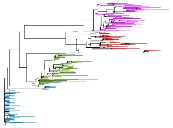
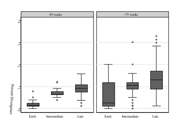
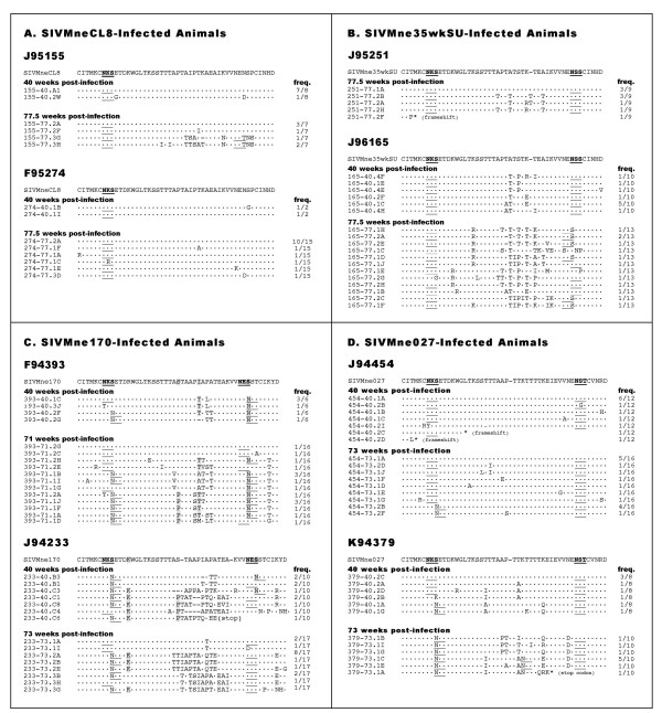
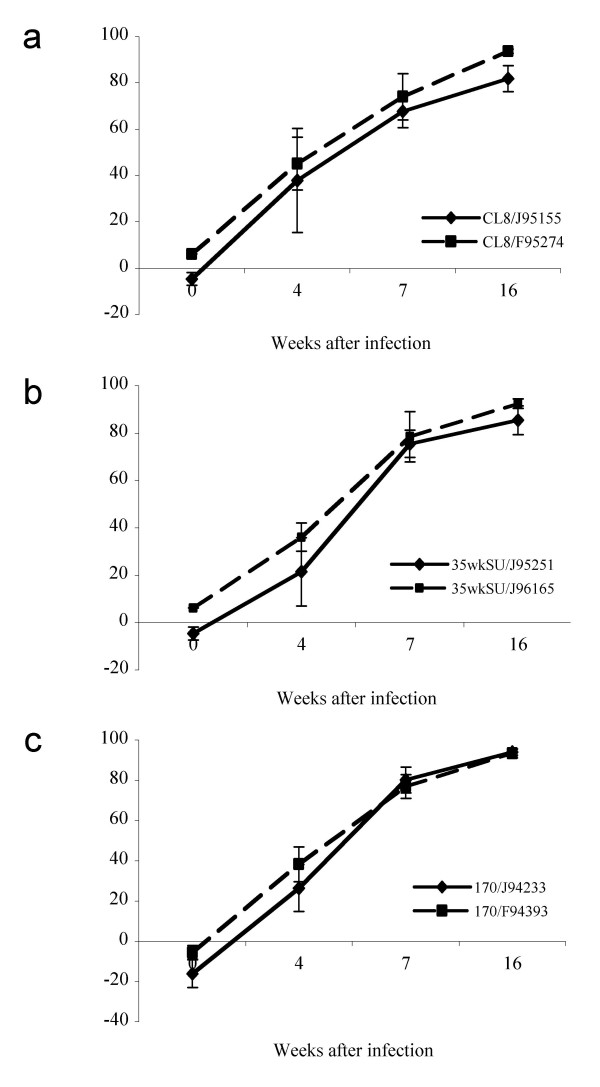
Background: Lentiviruses such as human and simian immunodeficiency viruses (HIV and SIV) undergo continual evolution in the host. Previous studies showed that the late-stage variants of SIV that evolve in one host replicate to significantly higher levels when transmitted to a new host. However, it is unknown whether HIVs or SIVs that have higher replication fitness are more genetically stable upon transmission to a new host. To begin to address this, we analyzed the envelope sequence variation of viruses that evolved in animals infected with variants of SIVMne that had been cloned from an index animal at different stages of infection.

Results: We found that there was more evolution of envelope sequences from animals infected with the late-stage, highly replicating variants than in animals infected with the early-stage, lower replicating variant, despite the fact that the late virus had already diversified considerably from the early virus in the first host, prior to transmission. Many of the changes led to the addition or shift in potential-glycosylation sites-, and surprisingly, these changes emerged in some cases prior to the detection of neutralizing antibody responses, suggesting that other selection mechanisms may be important in driving virus evolution. Interestingly, these changes occurred after the development of antibody whose anti-viral function is dependent on Fc-Fcgamma receptor interactions.

Conclusion: SIV variants that had achieved high replication fitness and escape from neutralizing antibodies in one host continued to evolve upon transmission to a new host. Selection for viral variants with glycosylation and other envelope changes may have been driven by both neutralizing and Fcgamma receptor-mediated antibody activities.


Figures

Figure 1
Figure 1
Phylogenetic relationship of viral variants. A distance-based tree was created using all unique sequences from all animals at both 40 and approximately 75 weeks-post infection. Sequences from SIVMneCL8-infected animals are shown in blue, those from SIVMne35wkSU-infected animals are shown in green, and those from SIVMne170- and SIVMne027-infected animals are shown in red and pink respectively. The parental sequences are marked by a diamond of the respective color.
Figure 2
Figure 2
Percent divergence from the infecting variant. For each animal, genetic distances were calculated between each sequence and the infecting variant, and animals were grouped by infecting variant. Box plots show the divergence of sequences from animals infected with the early variant (SIVMneCL8), the intermediate variant (SIVMne35wkSU), and the late variants (SIVMne170 and SIVMne027), at 40 and ~75 weeks post-infection.
Figure 3
Figure 3
V1 sequence variants. Amino acid sequence data from the V1 region of envelope is shown for each animal at each time point analyzed. Each sequence represents a unique variant and the frequency with which it was observed is shown in the column to the right. The parental V1 sequence is shown at the top of each alignment, and the conserved amino acids in each variant sequence are shown as dots. Sites of potential N-linked glycosylation are underlined in each sequence, and positions of reversion to the amino acid found in SIVMneCL8 are highlighted in grey.
Figure 4
Figure 4
Neutralization IC50s of sera collected at various times after infection against the infecting virus. Each panel represents neutralization of macaques infected with the same virus: a) SIVMneCL8; b) SIVMne35wkSU; c) SIVMne170; d) SIVMne027. For each panel, the x axis shows the time when sera samples were collected. The y-axis shows the IC50 – the reciprocal dilution of sera required to inhibit infection by 50%. Neutralization IC50s measured by the TZM-bl cells are shown in solid lines. Neutralization IC50s measured in sMAGI cells are shown in dotted lines.
Figure 5
Figure 5
ADCVI antibody activity in plasma of infected animals. Each panel represents th ADCVI activity (% inhibition) in 1:100 dilutions of plasma from animals infected with the same virus: (a) SIVMneCL8; b) SIVMne35wkSU; c) SIVMne170. Data are means ± standard error of four measurements for each animal at each timepoint.

Similar articles

Cited by

References

    1. Overbaugh J, Bangham CR. Selection forces and constraints on retroviral sequence variation. Science. 2001;292:1106–1109. doi: 10.1126/science.1059128. - DOI - PubMed
    1. Shankarappa R, Margolick JB, Gange SJ, Rodrigo AG, Upchurch D, Farzadegan H, Gupta P, Rinaldo CR, Learn GH, He X, Huang XL, Mullins JI. Consistent viral evolutionary changes associated with the progression of human immunodeficiency virus type 1 infection. J Virol. 1999;73:10489–10502. - PMC - PubMed
    1. Burns DP, Desrosiers RC. Selection of genetic variants of simian immunodeficiency virus in persistently infected rhesus monkeys. J Virol. 1991;65:1843–1854. - PMC - PubMed
    1. Johnson PR, Hamm TE, Goldstein S, Kitov S, Hirsch VM. The genetic fate of molecularly cloned simian immunodeficiency virus in experimentally infected macaques. Virology. 1991;185:217–228. doi: 10.1016/0042-6822(91)90769-8. - DOI - PubMed
    1. Overbaugh J, Rudensey LM, Papenhausen MD, Benveniste RE, Morton WR. Variation in simian immunodeficiency virus env is confined to V1 and V4 during progression to simian AIDS. J Virol. 1991;65:7025–7031. - PMC - PubMed

Publication types

MeSH terms