. 2008 Aug;8(2):146-56.
doi: 10.1016/j.cmet.2008.07.002.

The hepcidin-binding site on ferroportin is evolutionarily conserved

Affiliations

The hepcidin-binding site on ferroportin is evolutionarily conserved

Ivana De Domenico et al. Cell Metab. 2008 Aug.

Retraction in

Abstract

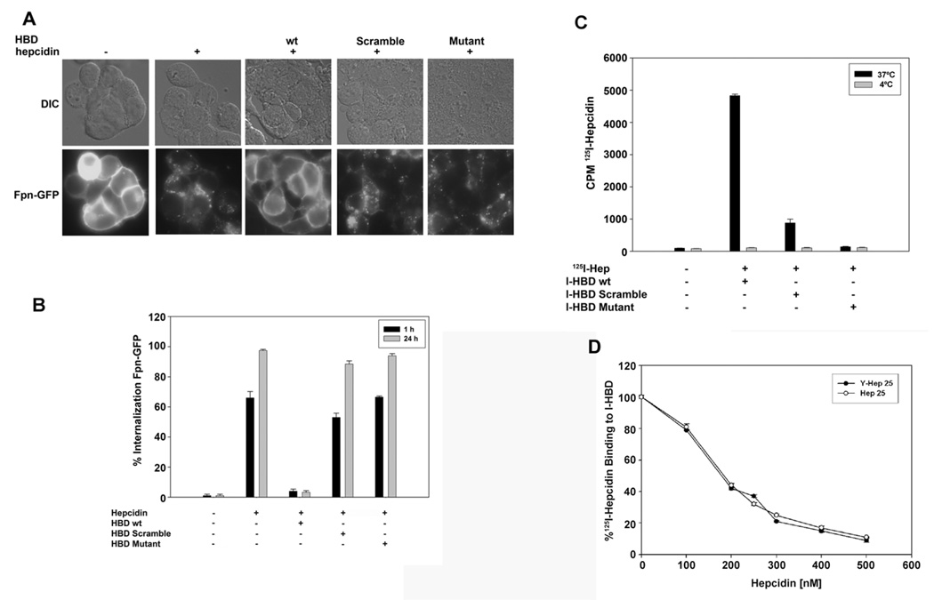
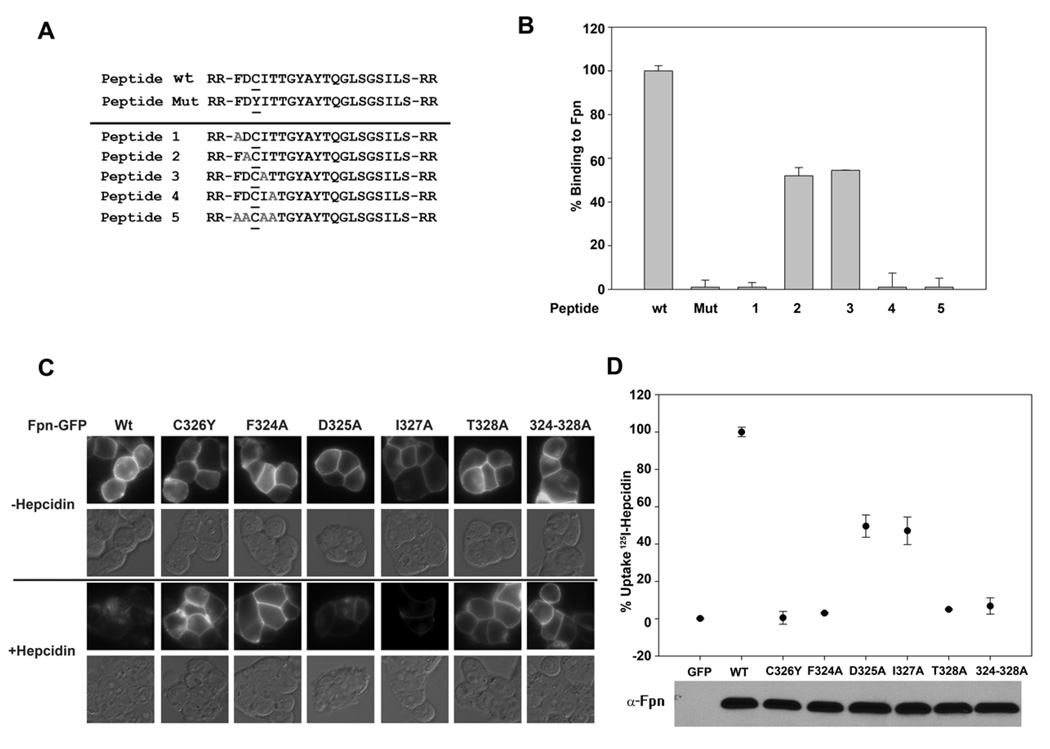
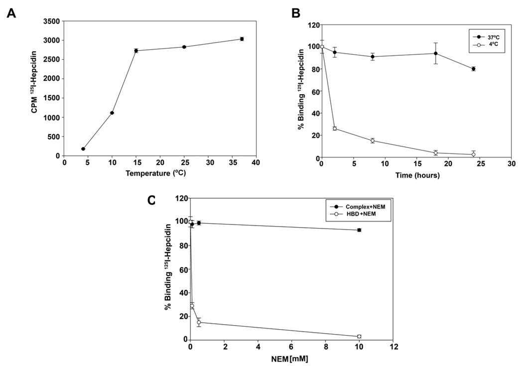
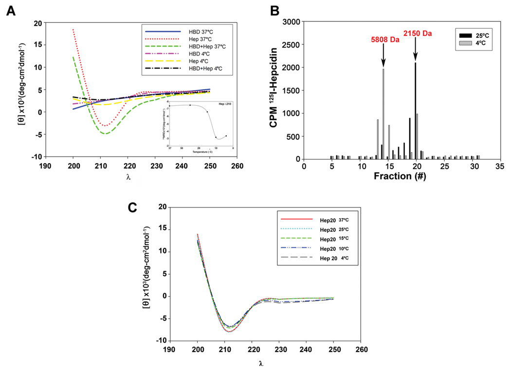
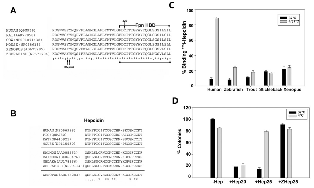
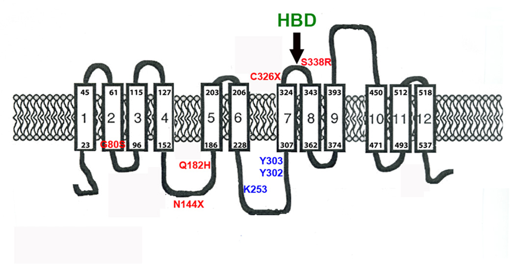
Mammalian iron homeostasis is regulated by the interaction of the liver-produced peptide hepcidin and its receptor, the iron transporter ferroportin. Hepcidin binds to ferroportin resulting in degradation of ferroportin and decreased cellular iron export. We identify the hepcidin-binding domain (HBD) on ferroportin and show that a synthetic 19 amino acid peptide corresponding to the HBD recapitulates the characteristics and specificity of hepcidin binding to cell-surface ferroportin. The binding of mammalian hepcidin to ferroportin or the HBD shows an unusual temperature dependency with an increased rate of dissociation at temperatures below 15 degrees C. The increased rate of dissociation is due to temperature- dependent changes in hepcidin structure. In contrast, hepcidin from poikilothermic vertebrates, such as fish or frogs, binds the HBD in a temperature-independent fashion. The affinity of hepcidin for the HBD permits a rapid, sensitive assay of hepcidin from all species and yields insights into the evolution of hepcidin.


Figures

Figure 1
Figure 1. HBD inhibits binding of hepcidin to Fpn
A. HEK293TFpn-GFP cells were incubated with or without hepcidin (1 µg/ml) or with hepcidin that had been pre-incubated either with HBD, scrambled-HBD or HBD with the C326Y mutation (HBD Mutant). (The number of the amino acid is based on its position in the full-length protein.) The HBD peptides and hepcidin were mixed at an equimolar ratio for two hrs at 37°C and then added to cells for one or 24 hrs. Fpn internalization at one hr was analyzed by epifluorescence. B. The percentage of cells showing internalized Fpn-GFP at one and 24 hrs was quantified. The data are reported as the standard error of the mean and were determined by counting 10 fields containing 20–30 cells/field. C. Peptides (HBD, scrambled-HBD or mutant (C326Y HBD)) were conjugated to agarose beads (I-HBD). 125I-hepcidin was added to I-HBD for 18 hrs at 37°C (black bars) or 4°C (grey bars). The beads were washed and the amount of 125I-hepcidin bound to beads was determined as counts per minute (cpm). D. Specified concentrations of hepcidin (Hep25) or hepcidin containing a tyrosine residue (M21Y, Y-Hep25) were added to I-HBD for one hr prior to the addition of 125I-hepcidin. The mixture was incubated for 18 hrs at 37°C and the amount of 125I-hepcidin bound to beads was determined. The data are expressed as the % 125I-hepcidin bound to I-HBD.
Figure 2
Figure 2. Effect of amino acid substitutions on the binding of hepcidin to the HBD and cell surface Fpn
A. HBDs containing amino acid substitutions (grey alanines A) were synthesized. B. The ability of modified HBD to bind 125I-hepcidin was assayed as described in Figure 1. C. Amino acid substitutions were generated in Fpn-GFP by site-specific mutagenesis as described in Experimental Procedures. Plasmids containing wild type or mutant Fpn-GFP were transiently transfected into HEK293T cells for 18–24 hrs. The transfected cells were incubated with hepcidin (4 µg/ml) for 4 hrs and internalization of Fpn-GFP was assayed by epifluorescence microscopy. D.125I-hepcidin was added to HEK293T cells expressing wt Fpn-GFP or mutant Fpn-GFP, and cell-associated radioactivity was measured. The expression of Fpn-GFP constructs was assessed by Western blot analysis using antibodies to Fpn with 25 µg of protein loaded per lane. The data are expressed as % uptake in which binding to cells expressing wild type Fpn-GFP was normalized as 100%.
Figure 3
Figure 3. Dissociation of mammalian hepcidin-HBD complexes is accelerated by low temperature
A. A sub-saturating concentration of 125I-hepcidin (200 nM) was incubated with I-HBD for 18 hrs at the specified temperatures and the amount of radioactivity bound to beads determined. B. 125I-hepcidin was added to I-HBD and a complex allowed to form at 37°C. The beads were washed, incubated with excess non-radioactive hepcidin at 37°C (closed circles) or 4°C (open circles) for the specified times and the amount of radioactivity bound to beads determined. The data are expressed as radioactivity at each time point relative to the amount bound at zero time. C. I-HBD was incubated with different concentrations of NEM for four hrs at 37°C. The beads were washed, I25I-hepcidin added and the amount of radioactivity bound to beads determined (open circles). Alternatively, the I-HBD-125I-hepcidin complex was allowed to form at 37°C. The beads were washed and then incubated with the specified concentrations of NEM for four hrs and radioactivity bound to the beads determined (closed circles). The data are expressed as the amount of radioactivity at each time point relative to the amount of radioactivity bound to beads prior to NEM treatment.
Figure 4
Figure 4. Temperature dependent changes in mammalian hepcidin structure
A. Human hepcidin (Hep), HBD or an equimolar mixture of HBD and hepcidin were analyzed by circular dichroism (CD) at 37°C and 4°C from wavelength (λ) 190 to 260. The insert shows the effect of temperature on the CD spectrum of hepcidin measured at 210 nm. B. 125I-hepcidin was applied to a G-25 column, equilibrated and eluted at 25°C (black bars) or 4°C (grey bars) and the amount of radioactivity in eluted fractions determined. The arrows represent the elution of molecular weight standards, insulin (5808 Da) and DBI (2150 Da). The elution of the standards was not affected by temperature. C. CD spectrum of Hep20 (100 µM) measured at different temperatures.
Figure 5
Figure 5. Alignment of vertebrate Fpn-HBD and hepcidin and the effect of temperature on hepcidin activity
A. Alignment of HBD sequence from homeothermic (warm blooded) and poikilothermic (cold-blooded) vertebrates. The arrows denote the location of the tyrosines that are phosphorylated upon hepcidin binding and cysteine 326 which when mutated leads to hepcidin resistance and ferroportin linked hemochromatosis. “*” represents identity and “:” represents similarity. The accession numbers are listed next to the sequence. B. Alignment of hepcidin sequence from mammals, fish and Xenopus. C. I-HBD was incubated with human or zebrafish hepcidin, or with serum from Salmo trutta (brown trout), Pungitius pungitius (Alaskan nine spine stickleback) or Xenopus laevis for 12 hrs at 37°C. The beads were washed three times and 125I-hepcidin was added for one hr at 37°C. The amount of radioactivity bound was determined and compared to that bound to I-HBD that had been incubated with 125I-hepcidin for one hr (black bars). An aliquot of I-HBD that had been incubated with hepcidin or sera were washed and then incubated at 4°C for 12 hrs. Following washing, 125I-hepcidin was added at 37°C and the amount of radioactivity determined after one hr (grey bars). D. E.coli, grown in LB broth were incubated with 30 µM of hepcidin (Hep25), Hep20 or zebrafish hepcidin (ZHep25) at 37°C (black bars) or 4°C (grey bars) for 4 hrs. Aliquots of bacteria were plated on LB and incubated at 37 °C for 12 hrs and the number of colonies determined. The data are expressed as the percent of colonies observed in the absence of hepcidin. The error bars represent standard error of the mean.
Figure 6
Figure 6. Determination of hepcidin concentrations in human and mouse sera
A. Samples (25 µL) from pooled human sera were incubated with I-HBD and a known concentration of 125I-hepcidin for 18 hrs at 37°C. The beads were then washed and the amount of radioactivity determined. Serum hepcidin levels were calculated relative to a standard curve constructed using chemically synthesized human hepcidin. For these and the data described below, each sample was measured in triplicate and the error bars are the standard error of the mean. B. Hepcidin levels were determined in sera obtained from normal individuals of defined age and gender or in serum obtained from a female patient diagnosed with juvenile hemochromatosis (JH) due to mutations in HJV gene as described in A. C. Hepcidin levels in sera obtained from wild type mice (C57BL/6) from mice homozygous (HAMP−/− ) or heterozygous (HAMP+/− ) for a targeted gene deletion in HAMP or mice that were homozygous for a targeted gene deletion in HFE were determined as described in A. D. C3H/HeJ mice were injected intraperitoneally with LPS, serum was obtained from the mice 12 hrs later and hepcidin concentration was determined as described in A. The data are expressed as percent of normal in which hepcidin levels in the absence of LPS is 100%.
Figure 7
Figure 7. Predicted topology of Fpn showing the position of mutations that lead to hepcidin resistance
The structure of Fpn is based on the study of Liu et al (Liu et al., 2005). We have added the locations of known human mutations that result in hepcidin resistant iron overload disease (S338R(Wallace et al., 2007), C326S(Sham et al., 2005), N144H(Njajou et al., 2002), Q182H(Hetet et al., 2003) and G80S(De Domenico et al., 2006a). We have also noted the positions of introduced amino acid substitutions that affect hepcidin binding and Fpn internalization (Y302, Y303) and degradation (K253A)(De Domenico et al., 2007b).

Comment in

References

    1. Casale G, Bonora C, Migliavacca A, Zurita IE, de Nicola P. Serum ferritin and ageing. Age Ageing. 1981;10:119–122. - PubMed
    1. De Domenico I, McVey Ward D, Nemeth E, Ganz T, Corradini E, Ferrara F, Musci G, Pietrangelo A, Kaplan J. Molecular and clinical correlates in iron overload associated with mutations in ferroportin. Haematologica. 2006a;91:1092–1095. - PMC - PubMed
    1. De Domenico I, Ward DM, Kaplan J. Hepcidin regulation: ironing out the details. J Clin Invest. 2007a;117:1755–1758. - PMC - PubMed
    1. De Domenico I, Ward DM, Langelier C, Vaughn MB, Nemeth E, Sundquist WI, Ganz T, Musci G, Kaplan J. The molecular mechanism of hepcidin-mediated ferroportin down-regulation. Mol Biol Cell. 2007b;18:2569–2578. - PMC - PubMed
    1. De Domenico I, Ward DM, Musci G, Kaplan J. Iron overload due to mutations in ferroportin. Haematologica. 2006b;91:92–95. - PMC - PubMed

Publication types

MeSH terms