. 2009 Sep;13(9B):3632-43.
doi: 10.1111/j.1582-4934.2008.00453.x. Epub 2008 Aug 4.

SIRT1 confers protection against UVB- and H2O2-induced cell death via modulation of p53 and JNK in cultured skin keratinocytes

Affiliations

SIRT1 confers protection against UVB- and H2O2-induced cell death via modulation of p53 and JNK in cultured skin keratinocytes

Cong Cao et al. J Cell Mol Med. 2009 Sep.

Abstract

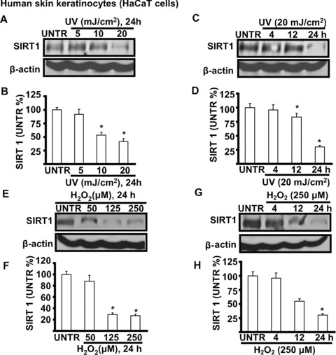
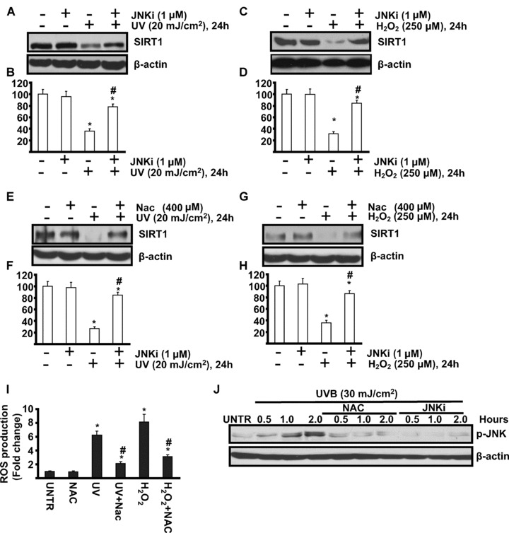
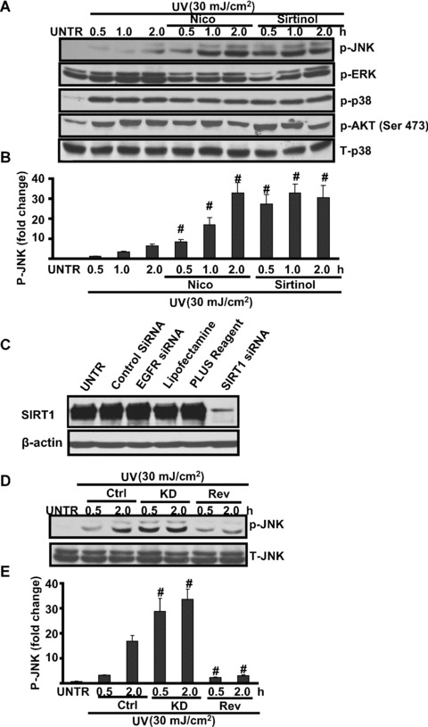
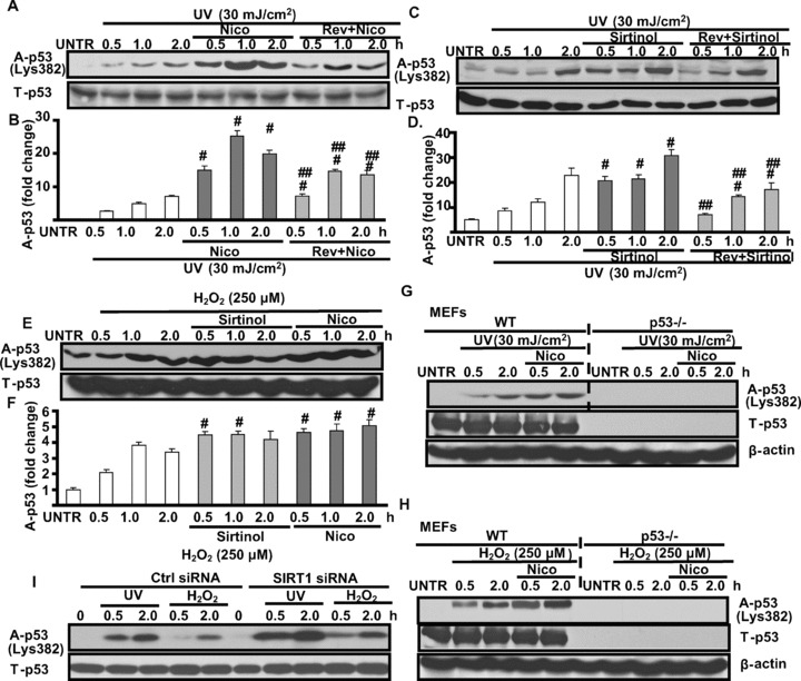
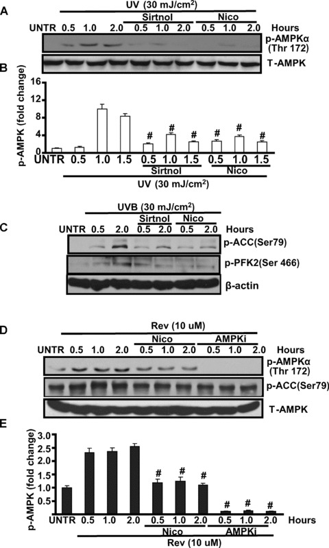
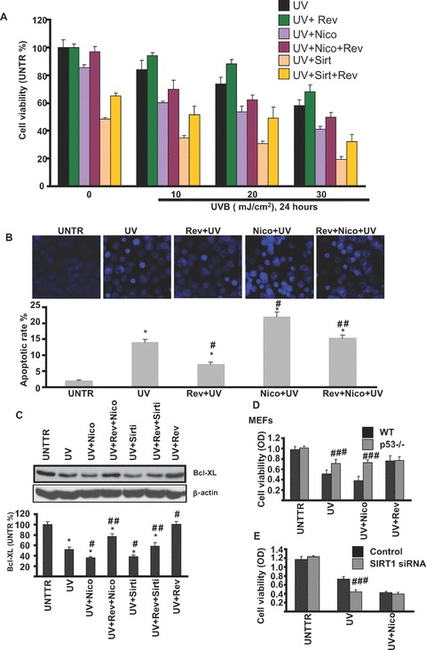
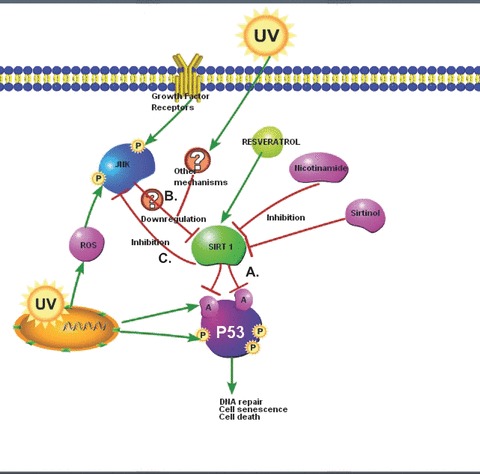
SIRT1 is a member of a highly conserved gene family (sirtuins) encoding nicotinamide adenine dinucleotide (NAD)(+)-dependent deacetylases, originally found to deacetylate histones leading to increased DNA stability and prolonged survival in yeast and higher organisms, including mammals. SIRT1 has been found to function as a deacetylase for numerous protein targets involved in various cellular pathways, including stress responses, apoptosis and axonal degeneration. However, the role of SIRT1 in ultraviolet (UV) signalling pathways remains unknown. Using cell culture and Western blot analysis in this study we found that SIRT1 is expressed in cultured human skin keratinocytes. Both UV radiation and H(2)O(2), two major inducers of skin cell damage, down-regulate SIRT1 in a time- and dose-dependent manner. We observed that reactive oxygen species-mediated JNK activation is involved in this SIRT1 down-regulation. SIRT1 activator, resveratrol, which has been considered as an important antioxidant, protects against UV- and H(2)O(2)-induced cell death, whereas SIRT inhibitors such as sirtinol and nicotinamide enhance cell death. Activation of SIRT1 negatively regulates UV- and H(2)O(2)-induced p53 acetylation, because nicotinamide and sirtinol as well as SIRT1 siRNA enhance UV- and H(2)O(2)-induced p53 acetylation, whereas SIRT1 activator resveratrol inhibits it. We also found that SIRT1 is involved in UV-induced AMP-activated protein kinase (AMPK) and downstream acetyl-CoA carboxylase (ACC), phosphofructose kinase-2 (PFK-2) phosphorylation. Collectively, our data provide new insights into understanding of the molecular mechanisms of UV-induced skin aging, suggesting that SIRT1 activators such as resveratrol could serve as new anti-skin aging agents.


Figures

Figure 1
Figure 1
UV and H2O2 down-regulate SIRT1 expression in cultured skin keratinocytes. HaCaT cells were treated with different doses of UV (5, 10 and 20 mJ/cm2) (A and B), cells then incubated in basic medium (DMEM) for 24 hrs or treated with 20 of mJ/cm2 UV and incubated in DMEM for different time-points (4, 12 and 24 hrs) (C and D), SIRT1 and β-actin were detected by Western blot. HaCaT cells were treated with different doses of H2O2 (50, 125 and 250 μM) for 24 hrs (E and F) or treated with 250 μM of H2O2 for different time-points (4, 12 and 24 hrs), SIRT1 and β-actin were detected by Western blot (G and H). The data in figures represent mean ± S.E. of three independent experiments. The symbol ‘*’ means P < 0.05 with untreated group (lane 1).
Figure 2
Figure 2
ROS-mediated JNK activation is involved in UV- and H2O2-induced SIRT1 down-regulation. HaCaT cells were pre-treated with JNK inhibitor (SP 600125, 1 μM, or JNKi) for 1 hr, followed by 20 mJ/cm2 UV radiation (A and B) or 250 μM of H2O2 (C and D) and incubated for 24 hrs, SIRT1 and β-actin expression were detected by Western blot. HaCaT cells were pre-treated with anti-oxidant NAC (n-acetyl-l-cysteine) (NAC, 400 μM) for 1 hr, followed by 20 mJ/cm2 UV radiation (E and F) or 250 μM of H2O2 (G and H) and incubated for 24 hrs, SIRT1 and β-actin expression were detected by Western blot. HaCaT cells were pre-treated with NAC for 1 hr, followed by 20 mJ/cm2 of UV radiation or 250 μM of H2O2 and incubated for 1 hr (I), ROS production was detected by FACS as described in methods. HaCaT cells were pre-treated with NAC (400 μM) or JNK inhibitor (SP 600125, 1 μM, or JNKi) for indicated time-points, p-JNK and β-actin were detected by Western blot. The data in figures represent mean ± S.E. of three independent experiments. The symbol ‘*’ means P < 0.05 with untreated group. The symbol ‘#’ means P < 0.05 with UV- or H2O2-treated group.
Figure 3
Figure 3
SIRT1 regulates UV-induced JNK activation. HaCaT cells were pre-treated with nicotinamide (Nico, 10 mM) or sirtinol (2 mM) for 1 hr, followed by UV radiation (25 mJ/cm2) and then incubated in DMEM for 0.5, 1.0 and 2.0 hrs, p-JNK, p-ERK, p-p38, p-AKT (473) and T-p38 were detected by Western blot (A) and JNK phosphorylation was quantified in (B). HaCaT cells with or without SIRT1 siRNA were pre-treated with resveratrol (Rev, 10 μM) for 1 hr, followed by UV radiation for indicated time-points, p-JNK and T-JNK were detected by Western blot. HaCaT cells were treated with 150 nM of SIRT1 siRNA or controls for 48 hrs, SIRT1 and β-actin were detected by Western blot (C). (D) Control or SIRT1 siRNA (150 nM) pre-treated cells were treated with UV radiation (25 mJ/cm2) for indicated time along with or without resveratrol (Rev, 10 μM, 1 hr prior to UV radiation), p-JNK and T-JNK were detected by Western blot, JNK phosphorylation was quantified in (E). The data in figures represent mean ± S.E. of three independent experiments. The symbol ‘#’ means P < 0.05 with UV-treated group.
Figure 4
Figure 4
SIRT1 negatively regulates UV- and H2O2-induced p53 acetylation. HaCaT cells were pre-treated with nicotinamide (Nico, 10 mM) or resveratrol (Rev, 10 μM) plus Nico for 1 hr, followed by UV radiation (25 mJ/cm2) and incubated for 0.5, 1.0 and 2.0 hrs, acelyated p53 and T-p53 were detected by Western blot (A and B). HaCaT cells were also pre-treated with sirtinol (2 mM) or Rev plus sirtinol for 1 hr, followed by UV radiation for indicated time, acelyated p53 and T-p53 were detected by Western blot (C and D). HaCaT cells were pre-treated with Nico or sirtinol for 1 hr, followed by H2O2 (250 μM), acelyated p-53 and T-p53 were detected by Western blot (E and F). Wild-type and p53 knockout MEFs were pre-treated with Nico for 1 hr, followed by UV (G) or H2O2 (H), for indicated time-points, acetylated p53 and T-p53 were detected by Western blot. Control and SIRT1 siRNA treated HaCaT cells were treated 30 mJ/cm2 of UV radiation or 250 μM of H2O2 for indicated time-points, acetylated p53 and T-p53 were detected by Western blot (I). The data in figures represent mean ± S.E. of three independent experiments. The symbol ‘#’ means P < 0.05 with UV- or H2O2-treated group, the symbol ‘##’ means P < 0.05 with UV plus Nico or sirtinol treated group.
Figure 5
Figure 5
SIRT1 positively regulates AMPK activation in cultured skin keratinocytes. HaCaT cells were pre-treated with sirtinol (2 mM) or nicotinamide (Nico, 10 mM) for 1 hr, followed by 20 mJ/cm2 of UV for indicated time, p-AMPK (Thr 172) and total-AMPK activation were detected by Western blot (A and B). HaCaT cells were pre-treated with sirtinol (2 mM) or nicotinamide (Nico, 10 mM) for 1 hr, followed by 20 mJ/cm2 of UV and incubated for 0.5 and 2.0 hrs, p-ACC (Ser 79), p-PFK-2 (Ser 466) and β-actin were detected by Western blot (C), p-ACC was quantified in (D). HaCaT cells were treated with nicotinamide (Nico, 10 mM) or AMPK inhibitor Compound C (AMPKi, 10 μM) for 0.5, 2.0 hrs, followed by resveratrol (Rev, 10 μM) treatment for 0.5, 1.0 and 2.0 hrs, p-AMPK (Thr 172), p-ACC (Ser 79) and T-AMPK were detected by Western blot (E and F). The data in figures represent mean ± S.E. of three independent experiments. The symbol ‘#’ means P < 0.05 with UV- or H2O2-treated group.
Figure 6
Figure 6
SIRT1 protects against UV-induced skin cell damage. HaCaT cells were pre-treated with resveratrol (Rev, 10 μM), sirtinol (2 mM), nicotinamide (Nico, 10 mM), Rev + Nico or Rev + sirtinol for 1 hr, followed by 10, 20 or 30 mJ/cm2 of UV radiation and incubated in DMEM for 24 hrs, cell viability was detected by MTT assay (A). HaCaT cells were pre-treated with nicotinamide, resveratrol, or Rev + Nico for 1 hr, followed by 20 mJ/cm2 of UV radiation and incubation in DMEM for 24 hrs, cell apoptosis was detected by Hoechst assay (B). HaCaT cells were treated with indicated treatments for 24 hrs, Bcl-xl and β-actin were detected by Western blot (C). Wild-type and p53 knockout MEFs were treated with UV, UV + Nico or UV + Rev and incubated in DMEM for 24 hrs, cell viability was detected by MTT assay (D). Control or SIRT1 siRNA pre-treated cells were treated with UV (20 mJ/cm2) or UV + Nico (Nico, 10 mM) for 24 hrs, cell viability were detected by MTT assay (E). The data in figures represent mean ± S.E. of three independent experiments. The symbol ‘#’ means P < 0.05 with UV- or H2O2-treated group, the symbol ‘##’ means P < 0.05 with UV plus Nico or sirtinol treated group, the symbol ‘####’ means P < 0.05 with p53 knockout or SIRT1 siRNA group.
Figure 7
Figure 7
Proposed cell signalling pathways involving SIRT1 in response to UV radiation. (A) SIRT1 negatively regulates UV- and H2O2-induced p53 acetylation. (B) ROS-mediated JNK activation is involved in UV-and H2O2-induced SIRT1 down-regulation. (C) SIRT1 inhibits UV-induced JNK activation.

Similar articles

Cited by

References

    1. Kaeberlein M, McVey M, Guarente L. The SIR2/3/4 complex and SIR2 alone promote longevity in Saccharomyces cerevisiae by two different mechanisms. Genes Dev. 1999;13:2570–80. - PMC - PubMed
    1. Vaziri H, Dessain SK, Ng Eaton E, et al. hSIR2(SIRT1) functions as an NAD-dependent p53 deacetylase. Cell. 2001;107:149–59. - PubMed
    1. Luo J, Nikolaev AY, Imai S, et al. Negative control of p53 by Sir2alpha promotes cell survival under stress. Cell. 2001;107:137–48. - PubMed
    1. Bouras T, Fu M, Sauve AA, et al. SIRT1 deacetylation and repression of p300 involves lysine residues 1020/1024 within the cell cycle regulatory domain 1. J Biol Chem. 2005;280:10264–76. - PubMed
    1. Brunet A, Bonni A, Zigmond MJ, et al. Akt promotes cell survival by phosphorylating and inhibiting a Forkhead transcription factor. Cell. 1999;96:857–68. - PubMed

Publication types