. 2008 Sep;69(6):1560-74.
doi: 10.1111/j.1365-2958.2008.06387.x. Epub 2008 Aug 4.

RloC: a wobble nucleotide-excising and zinc-responsive bacterial tRNase

Affiliations

RloC: a wobble nucleotide-excising and zinc-responsive bacterial tRNase

Elena Davidov et al. Mol Microbiol. 2008 Sep.

Abstract

The conserved bacterial protein RloC, a distant homologue of the tRNA(Lys) anticodon nuclease (ACNase) PrrC, is shown here to act as a wobble nucleotide-excising and Zn(++)-responsive tRNase. The more familiar PrrC is silenced by a genetically linked type I DNA restriction-modification (R-M) enzyme, activated by a phage anti-DNA restriction factor and counteracted by phage tRNA repair enzymes. RloC shares PrrC's ABC ATPase motifs and catalytic ACNase triad but features a distinct zinc-hook/coiled-coil insert that renders its ATPase domain similar to Rad50 and related DNA repair proteins. Geobacillus kaustophilus RloC expressed in Escherichia coli exhibited ACNase activity that differed from PrrC's in substrate preference and ability to excise the wobble nucleotide. The latter specificity could impede reversal by phage tRNA repair enzymes and account perhaps for RloC's more frequent occurrence. Mutagenesis and functional assays confirmed RloC's catalytic triad assignment and implicated its zinc hook in regulating the ACNase function. Unlike PrrC, RloC is rarely linked to a type I R-M system but other genomic attributes suggest their possible interaction in trans. As DNA damage alleviates type I DNA restriction, we further propose that these related perturbations prompt RloC to disable translation and thus ward off phage escaping DNA restriction during the recovery from DNA damage.


Figures

Fig. 1
Fig. 1
Functional organization of PrrC and RloC. A. Domain alignment. The ATPase domain of PrrC and ATPase head domain of RloC are indicated by pink rectangles, the ACNase domains by green rectangles; predicted α-helical regions flanking the CXXC zinc-hook motif thought to form an antiparallel coiled-coil bundle (CC) (Hopfner et al., 2002) are in grey, the gap containing the CXXC motif is in pink and the motif itself is in yellow. Dashed lines connect motifs shared by PrrC and RloC including the Walker A (P-loop), ABC signature, Walker B/D-loop, linchpin histidine/switch region (Moody and Thomas, 2005) and catalytic ACNase triad (Blanga-Kanfi et al., 2006). The A-loop [base specificity motif of typical ABC ATPases (Ambudkar et al., 2006)] is missing from PrrC whereas the PrrC Box (Blanga-Kanfi et al., 2006) and the region implicated in tRNA recognition have been described only in PrrC (Klaiman et al., 2007). B. COILED-COIL predictions of RloC orthologues encoded by the indicated bacterial strains. The arrow points at the position of the CXXC motif. C. Alignment of selected E. coli PrrC and G. kaustophilus HTA426 RloC sequences containing shared functional motifs (highlighted).
Fig. 2
Fig. 2
Expression of wild-type and mutated G. kaustophilus RloC forms in E. coli. A. RloC's expression is limited. E. coli Rosetta encoding wild-type G. kaustophilus RloC (lanes 1, 2), wild-type PrrC (lanes 3, 4) or the inactive PrrC-H356A mutant (lanes 5–8) not induced (odd lanes) or induced with 100 μM IPTG (even lanes) were lysed, the cellular proteins separated by SDS-PAGE and the recombinant ACNase proteins monitored by immunoblotting using an anti-His6 monoclonal antibody. The extract with the inactive PrrC mutant H356A was diluted in lanes 6 and 7 100- or 10-fold respectively. B. In vivo ACNase activity and protein level of RloC catalytic triad and zinc-hook mutants. E. coli Rosetta cells encoding the indicated RloC alleles were analysed for RloC protein level and in vivo ACNase activity as detailed in Experimental procedures.
Fig. 3
Fig. 3
RloC-expressing cells manifest ACNase activity. A. Total RNA samples isolated from cells expressing PrrC (odd lanes) or RloC (even lanes) were separated by denaturing polyacrylamide gel electrophoresis as such (lanes 1, 2) or after being 5′-end-labelled by T4 Pnk (lanes 5, 6). The non-labelled RNA fractions were also further incubated with T4 Pnk and Rnl 1 (lanes 3, 4) and then ligated with Rnl1 (lanes 7, 8). The gel was then stained with ethidium bromide (lanes 1–4) or autoradiographed (lanes 5–8). 33mers are 5′-cleavage products generated by either ACNase. 43mers are 3′-cleavage products generated by PrrC, ∼42mers and ∼52mers 3′-cleavage products generated by RloC. Band a contains the ligated PrrC cleavage products, bands a* and b contain the RloC counterparts, and bands c represents presumable internally ligated (circular) cleavage products of either ACNase. B. Scheme describing the cleavage of the ACNase substrate, 5′-end-labelling of the 3′-cleavage product, the subsequent ligation and the release of the labelled nucleotides from the labelled fragments or ligated-back molecules by the indicated nucleases. C. 2D TLC of radiolabelled nucleotides released by nuclease P1 (panels I–VII) or RNase T2 (panels IX–XI) from the indicated labelled RNA preparations. The 5′-end-labelled 43mers, ∼42mers and ∼52mers (Fig. 4A, lanes 1, 2), their ligated-back derivatives of bands a, a* and b (lanes 3, 4) and the circularized forms of RloC's products (lane 3) were digested by nuclease P1 and the released radiolabelled nucleotides separated by 2D TLC, as indicated. 5′-NMP markers are shown in panel VIII. The 3′-NMPs released from the indicated ligated-back derivatives by RNase T2 were similarly separated. The identity of U8 (panels I, II) was ascertained by subsequent separation on PEI-cellulose TLC (not shown). X1–X5 indicate apparent modified or hypomodified nucleotides that were not identified. D. Identification of tRNA species cleaved by PrrC or RloC. The 5′-end-labelled 43mers generated by PrrC (Fig. 4A, lane 1) or RloC's ∼42mers (Fig. 4A, lane 2) were hybridized to dot blots containing antisense DNA oligonucleotides corresponding to the indicated E. coli tRNA species described by Jiang et al. (2001) and in Table S2.
Fig. 4
Fig. 4
In vitro cleavage of tRNALys by PrrC or RloC. A. Cleavage products of tRNALys generated by PrrC or RloC. The tRNALys substrate labelled at the 33p34 junction was incubated with PrrC (lanes 1, 2) or RloC (lanes 3, 4) alone (lanes 1, 3) or in the presence of T4 Pnk providing 3′-cyclic phosphodiesterase/monoesterase (CPD) activities (lanes 2, 4). The products were separated by denaturing gel electrophoresis as detailed in Experimental procedures. B. 3′-end analysis of 33p>, 34p> and 34OH. The indicated labelled products obtained by tRNALys digestion with RloC (A, lane 1) or RloC and T4 Pnk (lane 2) were further digested with nuclease P1 and separated by PEI-thin-layer chromatography. The digestion products were identified by their position relative to markers produced by digesting tRNALys labelled at the 33p34 junction with (i) nuclease P1 to yield labelled pU8, (ii) RNase T2 to yield labelled Up, (iii) PrrC followed by nuclease P1 to yield labelled pUp>. The identity of pU8p> derived from 34p> by nuclease P1 digestion was ascertained by the ability to dephosphorylate it into labelled pU8 by incubation with T4 Pnk. C. Time-course of tRNALys cleavage by RloC. D. Proportions of the 34p> and 33p> cleavage products of tRNALys generated during the incubation with RloC. Shown are scanned profiles of the regions containing 34p> and 33p> in the indicated lanes of (C). E. Constancy of the 34p> fraction accessible to CPD. The cleavage of tRNALys by RloC was performed in the presence of CPD added either at the onset of the RloC reaction with RloC or 10 min later, as indicated. F. Scheme of tRNALys cleavage by PrrC or RloC and subsequent analyses of the cleavage products. The asterisk indicates the radiolabelled phosphate at the PrrC cleavage junction. The original substrate is represented by its anticodon stem loop region with positions of the canonical U33, the wobble base formula image 
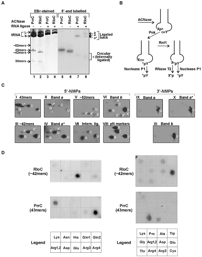
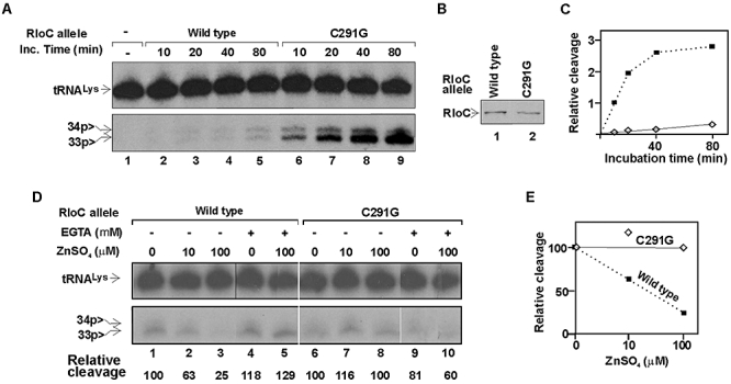
and second anticodon base U35 indicated. RloC cleaves the substrate initially 5′ to U35 yielding a 5′-fragment containing the radiolabel at the internal 33p34 position. The second cleavage by RloC at PrrC's site exposes the label to the phosphodiesterase/monoesterase activities of T4 Pnk (CPD) but can be pre-empted by prior 3′-dephosphorylation of 34p>. Nuclease P1 releases the indicated end groups from the various labelled products generated by RloC with or without CPD.
Fig. 5
Fig. 5
RloC and ACNase co-purify. A. Samples of the IMAC-purified wild-type RloC (lanes 1, 4), the ACNase-null E696A mutant (lanes 2, 5) or the zinc-hook mutant C291G (lanes 3, 6) derived from the same amount of total cell protein were separated by SDS-PAGE and monitored by immunoblotting using anti-His6-tag antibodies (lanes 1–3) or by silver staining (lanes 4–6). kDa, protein size markers. B. Samples of the indicated RloC alleles like those described in (A) were assayed for tRNALys cleavage activity as in Fig. 4.
Fig. 6
Fig. 6
Effects of Zn++ and EGTA on wild-type RloC or its zinc-hook mutant. A. Time-course of tRNALys cleavage by wild-type RloC or the zinc-hook mutant C291G. B. Western analysis of the S30 aliquots of the two RloC forms assayed in (A). C. Relative tRNALys cleavage versus incubation time with the indicated RloC allele. D. Effect of Zn++ and EGTA on the ACNase activity of the indicated RloC alleles. IMAC-purified fractions of the indicated RloC alleles were used in this assay. The reaction was performed under the standard conditions described in Experimental procedures and the incubation time was 30 min. E. Relative tRNALys cleavage activity versus zinc sulphate concentration.
Fig. 7
Fig. 7
G. kaustophilus rloC is linked to an hsdR relic. The schemes describing the region about G. kaustophilus rloC and its flanking hsdR relic and the complete hsd locus located elsewhere in the G. kaustophilus genome were adapted from the annotated genomic NCBI map (Takami et al., 2004). Numbers followed by nt indicate positions in the genomic map. The non-coding region separating locus tags GK0884-5 was found by blast (Altschul et al., 1997) to encode a C-terminal 568 aa portion of an HsdR species that was 98% identical in amino acid sequence with the matching portion of the hsdR gene of the complete locus (GK0346).

References

    1. Abdul-Jabbar M, Snyder L. Genetic and physiological studies of an Escherichia coli locus that restricts polynucleotide kinase and RNA ligase mutants of bacteriophage T4. J Virol. 1984;51:522–529. - PMC - PubMed
    1. Altschul SF, Madden TL, Schaffer AA, Zhang J, Zhang Z, Miller W, Lipman DJ. Gapped blast and psi-blast: a new generation of protein database search programs. Nucleic Acids Res. 1997;25:3389–3402. - PMC - PubMed
    1. Ambudkar SV, Kim IW, Xia D, Sauna ZE. The A-loop, a novel conserved aromatic acid subdomain upstream of the Walker A motif in ABC transporters, is critical for ATP binding. FEBS Lett. 2006;580:1049–1055. - PubMed
    1. Amitsur M, Levitz R, Kaufmann G. Bacteriophage T4 anticodon nuclease, polynucleotide kinase and RNA ligase reprocess the host lysine tRNA. EMBO J. 1987;6:2499–2503. - PMC - PubMed
    1. Amitsur M, Morad I, Kaufmann G. In vitro reconstitution of anticodon nuclease from components encoded by phage T4 and Escherichia coli CTr5X. EMBO J. 1989;8:2411–2415. - PMC - PubMed

Publication types