. 2008 Aug 6;16(8):1166-74.
doi: 10.1016/j.str.2008.04.012.

Structure of the E. coli DNA glycosylase AlkA bound to the ends of duplex DNA: a system for the structure determination of lesion-containing DNA

Affiliations

Structure of the E. coli DNA glycosylase AlkA bound to the ends of duplex DNA: a system for the structure determination of lesion-containing DNA

Brian R Bowman et al. Structure. .

Abstract

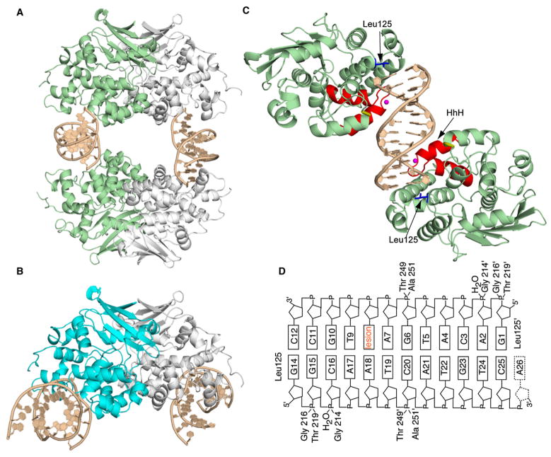
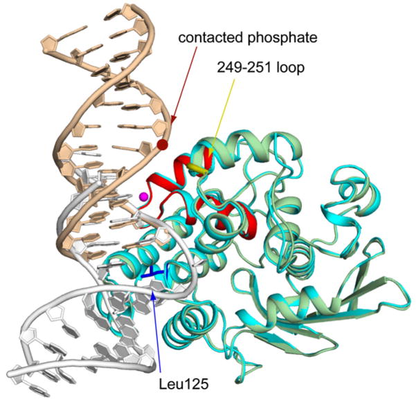
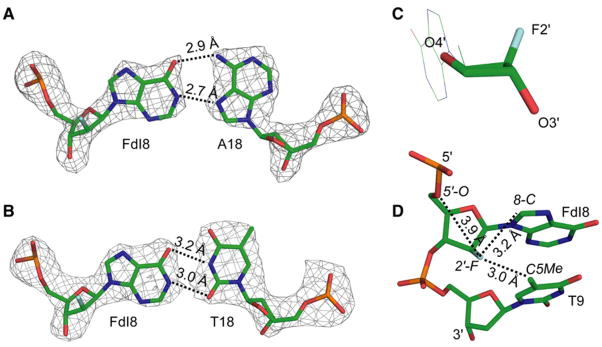
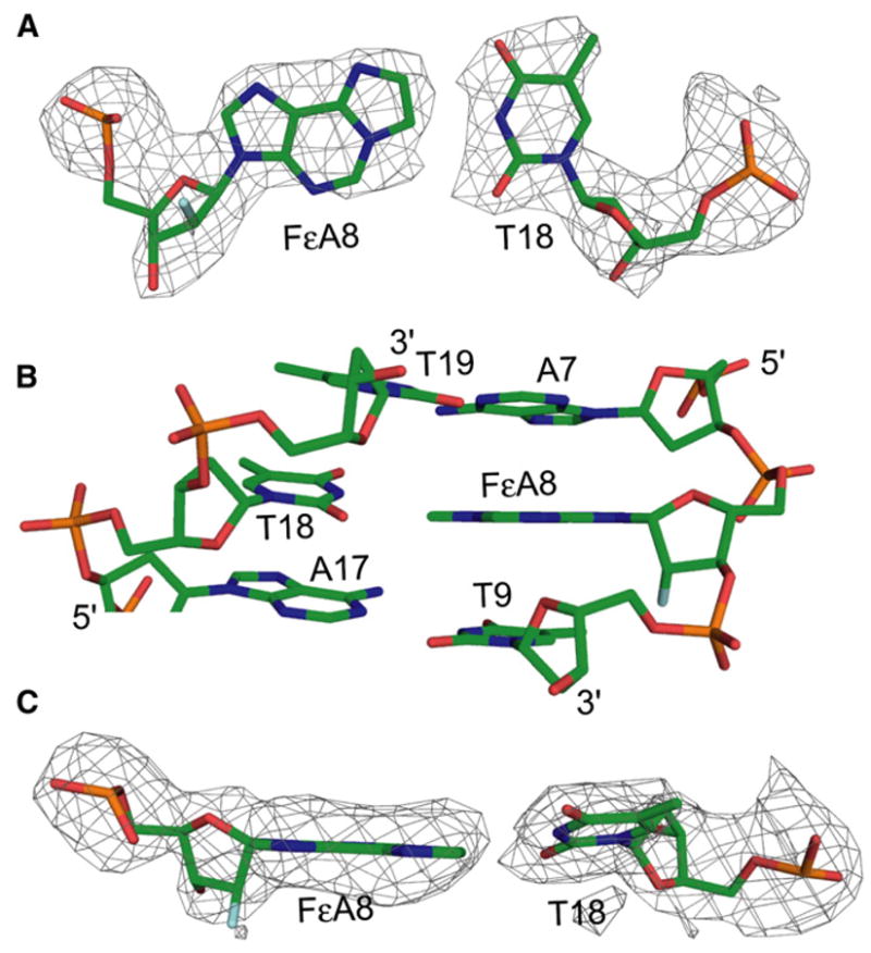
The constant attack on DNA by endogenous and exogenous agents gives rise to nucleobase modifications that cause mutations, which can lead to cancer. Visualizing the effects of these lesions on the structure of duplex DNA is key to understanding their biologic consequences. The most definitive method of obtaining such structures, X-ray crystallography, is troublesome to employ owing to the difficulty of obtaining diffraction-quality crystals of DNA. Here, we present a crystallization system that uses a protein, the DNA glycosylase AlkA, as a scaffold to mediate the crystallization of lesion-containing duplex DNA. We demonstrate the use of this system to facilitate the rapid structure determination of DNA containing the lesion 8-oxoguanine in several different sequence contexts, and also deoxyinosine and 1,N(6)-ethenoadenine, each stabilized as the corresponding 2'-flouro analog. The structures of 8-oxoguanine provide a correct atomic-level view of this important endogenous lesion in DNA.


Figures

Figure 1
Figure 1. Structure of the AlkA:DNA Host/Guest Complex
(A) Ribbon representation of the asymmetric unit in the AlkA:DNA host/guest complex (HGC) structure, with four AlkA monomers bookending two DNA duplexes. The two AlkA monomers in green are those that interact with the DNA duplex comprising chains G and H; this half of the structure is slightly more ordered than the other half and hence was used for the analyses throughout this work. (B) Ribbon representation of the AlkA azaribose lesion-recognition complex (Hollis et al., 2000). One of the AlkA monomers is colored cyan. Note the difference in the DNA-binding mode between (A) and (B). (C) Ribbon representation of the two AlkA monomers interacting with DNA chains G and H, (same as in [A]). Shown in red is the signature helix-hairpin-helix DNA-binding motif common to all members of the superfamily to which AlkA belongs. The loop colored in gold contains residues 249 and 251, which also participate in interactions with the DNA. The water molecule mediating protein:DNA interactions is colored in magenta, whereas the side chain of Leu125 is shown in blue. (D) Schematic representation of the AlkA:DNA interactions. The prime sign (′) represents residues interacting from the second AlkA protomer.
Figure 2
Figure 2. Cα Superposition of the AlkA Azaribose LRC Structure onto an AlkA/DNA Subunit from the AlkA HGC
The rmsd of the LRC (the protein is colored cyan as in Figure 1B; DNA colored white) onto the HGC subunit (colored as in Figure 1C) is 0.37 Å. The crimson dot denotes the position of the phosphate that hydrogen bonds with the protein backbone at positions 249 and 251 (yellow loop); this phosphate is the only element of the otherwise naked central portion of the DNA that is contacted by the protein. The major sites of protein:DNA interaction are colored as in Figure 1C.
Figure 3
Figure 3. Analysis of FdI:A and FdI:T Base Pairs
(A and B) Structure of FdI base paired with (A) adenine and (B) thymine. The nucleotides are represented as sticks; atoms are colored as follows: carbon, green; nitrogen, blue; oxygen, red; phosphate, orange; and fluorine, cyan. The Fo − Fc electron density map of the base pair is represented as a mesh contoured at 3σ. Black, dotted lines represent hydrogen bonds. (C) Effect of 2′-fluorine substitution on the deoxyribose sugar pucker of the FdI base. The color scheme is as in (A). (D) Stick representation of the FdI base and the neighboring base, T9, illustrating the microenvironment of the 2′-fluorine atom. Dotted lines indicate distances from the 2′-fluorine to the three nearest atoms. All representations are from the FdI:A structure.
Figure 4
Figure 4. Analysis of the FεA:T Base Pair
(A) Stick representation of the FεA:T base pair. The color scheme is the same as in Figure 3. The Fo − Fc electron density map of the base pair is represented as a mesh contoured at 3σ. (B) FεA:T and the neighboring base pairs. The color scheme is as in Figure 3. (C) The FεA:T base pair as viewed from the major groove, illustrating the propeller twist angle of T18. The color scheme and electron density map are as in (A).
Figure 5
Figure 5. B-Form DNA Containing oxoG:dA or oxoG:dC Base Pairs
(A–C) The oxoG:C base pairs (left column) with oxoG in the (A) one position, (B) six position, and (C) eight position within the DNA duplex. In the right column, G:C structures in the same positions within the DNA as the respective lesion are shown. (D) Structure of oxoG base paired with adenine. The color scheme, Fo − Fc electron density map and hydrogen bonding are represented as in Figure 3.

References

    1. Banerjee A, Verdine GL. A nucleobase lesion remodels the interaction of its normal neighbor in a DNA glycosylase complex. Proc Natl Acad Sci USA. 2006;103:15020–15025. - PMC - PubMed
    1. Banerjee A, Yang W, Karplus M, Verdine GL. Structure of a repair enzyme interrogating undamaged DNA elucidates recognition of damaged DNA. Nature. 2005;434:612–618. - PubMed
    1. Banerjee A, Santos WL, Verdine GL. Structure of a DNA glycosylase searching for lesions. Science. 2006;311:1153–1157. - PubMed
    1. Barrett TE, Savva R, Barlow T, Brown T, Jiricny J, Pearl LH. Structure of a DNA base-excision product resembling a cisplatin inter-strand adduct. Nat Struct Biol. 1998;5:697–701. - PubMed
    1. Barrett TE, Scharer OD, Savva R, Brown T, Jiricny J, Verdine GL, Pearl LH. Crystal structure of a thwarted mismatch glycosylase DNA repair complex. EMBO J. 1999;18:6599–6609. - PMC - PubMed

Publication types

LinkOut - more resources