. 2008 Aug 7;3(2):196-206.
doi: 10.1016/j.stem.2008.07.001.

NANOG is a direct target of TGFbeta/activin-mediated SMAD signaling in human ESCs

Affiliations

NANOG is a direct target of TGFbeta/activin-mediated SMAD signaling in human ESCs

Ren-He Xu et al. Cell Stem Cell. .

Abstract

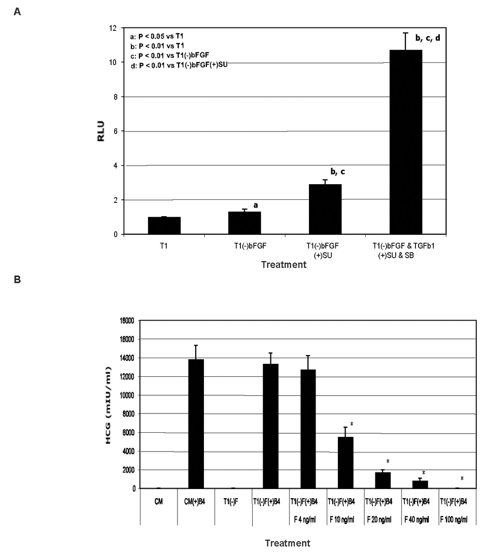
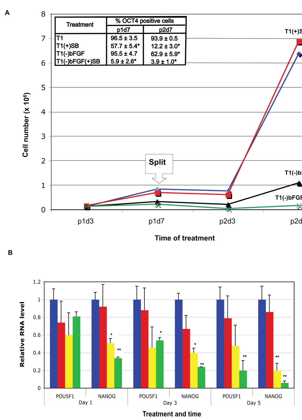
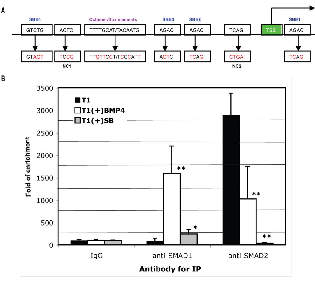
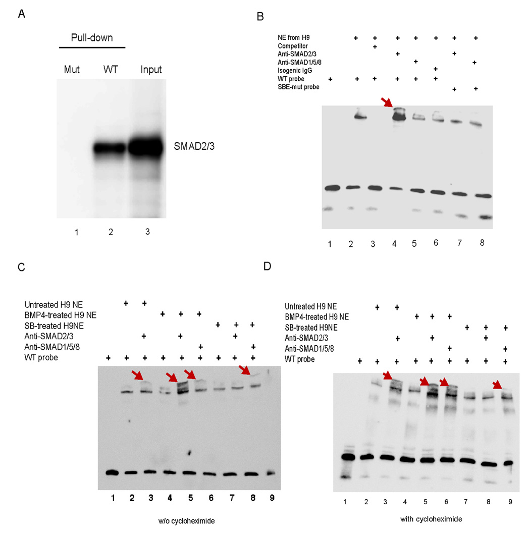
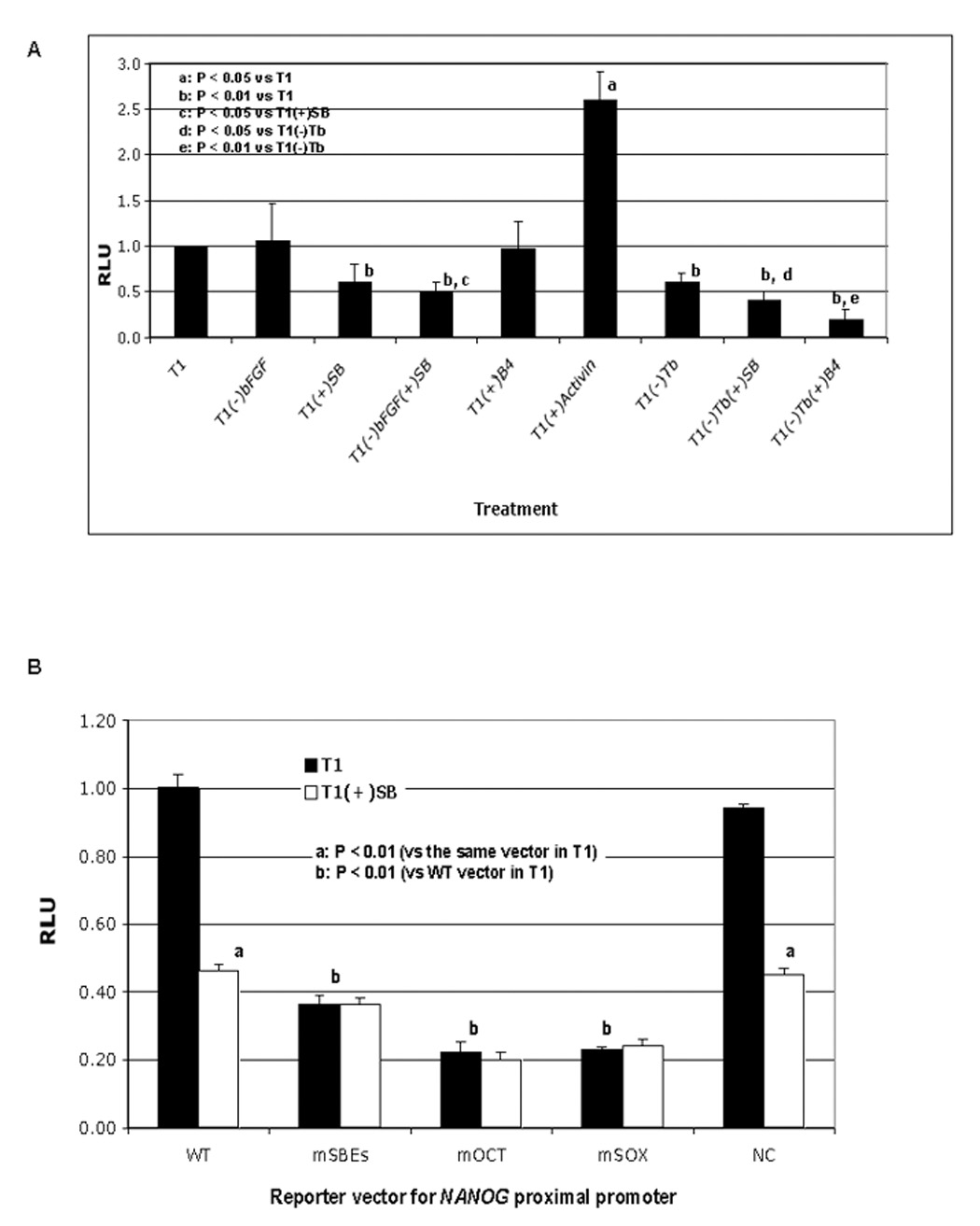
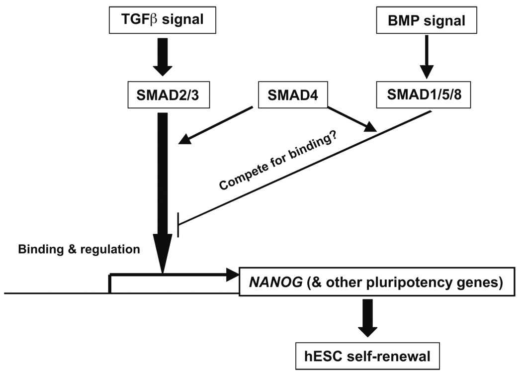
Self-renewal of human embryonic stem cells (ESCs) is promoted by FGF and TGFbeta/Activin signaling, and differentiation is promoted by BMP signaling, but how these signals regulate genes critical to the maintenance of pluripotency has been unclear. Using a defined medium, we show here that both TGFbeta and FGF signals synergize to inhibit BMP signaling; sustain expression of pluripotency-associated genes such as NANOG, OCT4, and SOX2; and promote long-term undifferentiated proliferation of human ESCs. We also show that both TGFbeta- and BMP-responsive SMADs can bind with the NANOG proximal promoter. NANOG promoter activity is enhanced by TGFbeta/Activin and FGF signaling and is decreased by BMP signaling. Mutation of putative SMAD binding elements reduces NANOG promoter activity to basal levels and makes NANOG unresponsive to BMP and TGFbeta signaling. These results suggest that direct binding of TGFbeta/Activin-responsive SMADs to the NANOG promoter plays an essential role in sustaining human ESC self-renewal.


Figures

Fig. 1
Fig. 1. Either FGF or TGFβ signaling antagonizes BMP signaling
A. Both FGF and TGFβ signals synergistically repress BMP signal in human ES cells in luciferase reporter assays. H9 cells were transfected with pID120-Luc and a trace amount of pRL-tk (for internal control) on day 1. Treatments of the cells started on day 2 with T1 medium (T1) or T1 minus or plus 100 ng/ml bFGF, 0.6 ng/ml TGFβ1, 10 µM SU5402 (SU, inhibitor of FGF receptors), and 10 µM SB431542 (SB, inhibitor of TGFβ receptors). The cells were lysed on day 3 for analysis of luciferase activity. B. bFGF inhibits HCG secretion from trophoblast differentiated from BMP4-treated human ES cells. H14 cells cultured in fibroblast-conditioned medium (CM) were rinsed with DMEM/F12 basal medium and switched to CM or T1 medium with or without various concentrations of bFGF (F) and 100 ng/ml BMP4 (B4) for 7 days with daily refreshment of the media. The spent media were collected on day 7, and assayed for HCG. Results are shown as mean ± standard deviation. *P < 0.01 compared to T1(−)bFGF(+)BMP4. SB has been previously been shown to inhibit the levels of phosphorylated Smad2 and Smad3 in human ES cells (James et al., 2005).
Fig. 2
Fig. 2. FGF and TGFβ synergize to sustain human ES cell self-renewal
A. bFGF and TGFβ effects on H1 cell growth and maintenance of OCT4 expression. 3.5 105 H1 cells were seeded in individual wells of 6-well plates in triplicate and cultured in T1 medium, T1 plus 10 µM SB431542 [T1(+)SB], T1 minus 100 ng/ml bFGF [T1(−)bFGF], or T1 minus 100 ng/ml bFGF plus 10 µM SB [T1(−)bFGF(+)SB]. The cells were split 1:3 on day 7 into the corresponding media and cultured for an additional 7 days. Total cell number per group was counted on days 3 and 7 of each passage (labeled as p1d3, p1d7, p2d3, and p2d7, respectively), and OCT4+ cell percentage determined by flow cytometry on p1d7 and p2d7. Results are shown as mean ± standard deviation. *P < 0.01 compared to T1. B. Expression of pluripotency genes in human ES cells treated with various growth factors or inhibitors. H9 cells were cultured in T1, T1(−)bFGF, T1(+)SB, or T1(−)bFGF(+) SB medium for 1, 3 or 5 days and followed by QPCR for expression of OCT4 and NANOG. *P < 0.05, **P < 0.01 compared to T1.
Fig. 3
Fig. 3. ChIP for SMADs binding to the NANOG proximal promoter
A. Schematic representation of NANOG proximal promoter region (not to scale) and mutagenesis strategy (see Fig. S4 for its complete sequence). TSS: transcription start site; SBE: SMAD binding element; NC: sequence mutated for negative control mutation; and red letters for mutated nucleotide residues. B. ChIP assay for SMAD binding to the NANOG proximal promoter. H1 cells were cultured in T1 medium, T1 plus 10 µM SB431542 [T1(+)SB], or T1 plus 100 ng/ml BMP4 [T1(+)BMP4] for 24 hours. The cells were harvested and processed for ChIP with isogenic IgG, anti-SMAD1/5/8, or anti-SMAD2/3 antibodies. The enrichment of the precipitated DNA by each of the antibodies versus the IgG was analyzed by QPCR using primers flanking the proximal promoter region. Results from two duplicated experiments are shown as fold of DNA enrichment. *P < 0.05, **P < 0.01 compared to T1.
Fig. 4
Fig. 4. EMSA for SMAD binding to the NANOG proximal promoter
A. DNA pull-down to verify probes for EMSA. A biotin-labeled probe of 80 bp was synthesized by PCR from the NANOG proximal promoter in the plasmid pNANOG-Luc for a wild-type probe (WT) or from the mutated promoter in pNANOG(mSBEs)-Luc for a mutated probe (SBE mut). Only the WT probe was able to pull down SMAD2/3 from the extract of H9 cells as verified by western blotting. B–D. EMSA on the nuclear extract (NE) from H9 cells untreated (B) or treated with 100 ng/ml BMP4 or 10 µM SB431542 for 3 h in the absence (C) or presence (D) of 10 µg/ml cycloheximide (Biomol, Plymouth Meeting, PA), a protein synthesis inhibitor. An unlabeled probe at 200-fold concentration of the labeled probe was used as a competitor, anti-SMAD2/3 or anti-SMAD1/5/8 antibody for super-shift, and isogenic IgG as a control.
Fig. 5
Fig. 5. NANOG proximal promoter activity in luciferase reporter assay
A. Regulation of NANOG promoter reporter activity by FGF, TGFβ, and BMP signaling. H9 cells were transfected with pNANOG-Luc and a trace amount of pGL4.70 (as an internal control) on day 1. Treatments of the cells started on day 2 with T1 medium, T1 minus a specific signal stimulator 100 ng/ml bFGF or 0.6 ng/ml TGFβ1 (Tb), plus 10 ng/ml Activin, 100 ng/ml BMP4 (B4) or 10 µM SB431542 (SB). After 24 hours of treatment, cells were harvested for analysis of luciferase activity. B. Effect of SBE mutations on pNANOG-Luc activity. H9 cells were transfected with wild-type pNANOG-Luc (WT), pNANOG(mSBEs)-Luc (mSBEs), pNANOG(mOCT)-Luc (mOCT), pNANOG(mSOX)-Luc (mSOX) or pNANOG(NC)-Luc (NC) on day 1 (all with pGL4.70), cultured in T1 alone or T1 plus SB starting on day 2, and harvested after 24 hours of treatment for analysis of luciferase activity.
Fig. 6
Fig. 6. NANOG overexpression bypasses the need for both TGFβ and FGF signaling to sustain human ES cell self-renewal
H1 cells transduced with lentiviral particles containing NANOG or EGFP and selected by puromycin were split to plates with T1 medium. Next day, the medium was replaced with T1, T1 minus TGFβ1 plus 10 µM SB431542 [T1(−)TGFβ1(+)SB], T1 minus bFGF plus 10 µM SU5402 [T1(−)bFGF(+)SU], or T1(−)TGFβ1(+)SB(−)bFGF(+)SU. After 5-day treatment with these media, the cells were fixed and processed for immunostaining for OCT4 expression. SU5402 has previously been shown to inhibit the level of phosphorylated ERK in human ES cells (Kang et al., 2005).
Fig. 7
Fig. 7. Model of SMAD regulation of NANOG transcription in human ES cells
Arrows represent induction and hammer-ended lines represent inhibition.

Comment in

References

    1. Amit M, Carpenter MK, Inokuma MS, Chiu CP, Harris CP, Waknitz MA, Itskovitz-Eldor J, Thomson JA. Clonally derived human embryonic stem cell lines maintain pluripotency and proliferative potential for prolonged periods of culture. Dev Biol. 2000;227:271–278. - PubMed
    1. Amit M, Shariki C, Margulets V, Itskovitz-Eldor J. Feeder layer- and serum-free culture of human embryonic stem cells. Biol Reprod. 2004;70:837–845. - PubMed
    1. Beattie GM, Lopez AD, Bucay N, Hinton A, Firpo MT, King CC, Hayek A. Activin A maintains pluripotency of human embryonic stem cells in the absence of feeder layers. Stem Cells. 2002;23:489–495. - PubMed
    1. Besser D. Expression of nodal, lefty-a, and lefty-B in undifferentiated human embryonic stem cells requires activation of Smad2/3. J Biol Chem. 2004;279:45076–45084. - PubMed
    1. Boyer LA, Lee TI, Cole MF, Johnstone SE, Levine SS, Zucker JP, Guenther MG, Kumar RM, Murray HL, Jenner RG, et al. Core transcriptional regulatory circuitry in human embryonic stem cells. Cell. 2005;122:947–956. - PMC - PubMed

Publication types

MeSH terms