Deiodinase-mediated thyroid hormone inactivation minimizes thyroid hormone signaling in the early development of fetal skeleton
- PMID: 18682303
- PMCID: PMC4683160
- DOI: 10.1016/j.bone.2008.06.020
Deiodinase-mediated thyroid hormone inactivation minimizes thyroid hormone signaling in the early development of fetal skeleton
Abstract
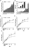
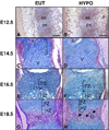
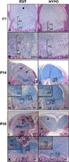
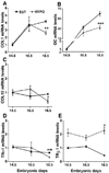
Thyroid hormone (TH) plays a key role on post-natal bone development and metabolism, while its relevance during fetal bone development is uncertain. To study this, pregnant mice were made hypothyroid and fetuses harvested at embryonic days (E) 12.5, 14.5, 16.5 and 18.5. Despite a marked reduction in fetal tissue concentration of both T4 and T3, bone development, as assessed at the distal epiphyseal growth plate of the femur and vertebra, was largely preserved up to E16.5. Only at E18.5, the hypothyroid fetuses exhibited a reduction in femoral type I and type X collagen and osteocalcin mRNA levels, in the length and area of the proliferative and hypertrophic zones, in the number of chondrocytes per proliferative column, and in the number of hypertrophic chondrocytes, in addition to a slight delay in endochondral and intramembranous ossification. This suggests that up to E16.5, thyroid hormone signaling in bone is kept to a minimum. In fact, measuring the expression level of the activating and inactivating iodothyronine deiodinases (D2 and D3) helped understand how this is achieved. D3 mRNA was readily detected as early as E14.5 and its expression decreased markedly ( approximately 10-fold) at E18.5, and even more at 14 days after birth (P14). In contrast, D2 mRNA expression increased significantly by E18.5 and markedly ( approximately 2.5-fold) by P14. The reciprocal expression levels of D2 and D3 genes during early bone development along with the absence of a hypothyroidism-induced bone phenotype at this time suggest that coordinated reciprocal deiodinase expression keeps thyroid hormone signaling in bone to very low levels at this early stage of bone development.
Figures





Similar articles
-
Thyroid hormones promote chondrocyte differentiation in mouse ATDC5 cells and stimulate endochondral ossification in fetal mouse tibias through iodothyronine deiodinases in the growth plate.J Bone Miner Res. 2002 Mar;17(3):443-54. doi: 10.1359/jbmr.2002.17.3.443. J Bone Miner Res. 2002. PMID: 11874236
-
The effect of 3,5,3'-triiodothyronine supplementation on zebrafish (Danio rerio) embryonic development and expression of iodothyronine deiodinases and thyroid hormone receptors.Gen Comp Endocrinol. 2007 Jun-Jul;152(2-3):206-14. doi: 10.1016/j.ygcen.2007.02.020. Epub 2007 Feb 27. Gen Comp Endocrinol. 2007. PMID: 17418841
-
The effect of diet-induced weight-loss on subcutaneous adipose tissue expression of genes associated with thyroid hormone action.Clin Nutr. 2024 Dec;43(12):245-250. doi: 10.1016/j.clnu.2024.11.006. Epub 2024 Nov 6. Clin Nutr. 2024. PMID: 39527910
-
Deiodinases and the Metabolic Code for Thyroid Hormone Action.Endocrinology. 2021 Aug 1;162(8):bqab059. doi: 10.1210/endocr/bqab059. Endocrinology. 2021. PMID: 33720335 Free PMC article. Review.
-
Role of the type 2 iodothyronine deiodinase (D2) in the control of thyroid hormone signaling.Biochim Biophys Acta. 2013 Jul;1830(7):3956-64. doi: 10.1016/j.bbagen.2012.08.019. Epub 2012 Aug 29. Biochim Biophys Acta. 2013. PMID: 22967761 Free PMC article. Review.
Cited by
-
Hormone-regulated expression and distribution of versican in mouse uterine tissues.Reprod Biol Endocrinol. 2009 Jun 5;7:60. doi: 10.1186/1477-7827-7-60. Reprod Biol Endocrinol. 2009. PMID: 19500372 Free PMC article.
-
Modulation of small leucine-rich proteoglycans (SLRPs) expression in the mouse uterus by estradiol and progesterone.Reprod Biol Endocrinol. 2011 Feb 4;9:22. doi: 10.1186/1477-7827-9-22. Reprod Biol Endocrinol. 2011. PMID: 21294898 Free PMC article.
-
Type 2 deiodinase at the crossroads of thyroid hormone action.Int J Biochem Cell Biol. 2011 Oct;43(10):1432-41. doi: 10.1016/j.biocel.2011.05.016. Epub 2011 Jun 12. Int J Biochem Cell Biol. 2011. PMID: 21679772 Free PMC article. Review.
-
Role of Thyroid Hormones in Skeletal Development and Bone Maintenance.Endocr Rev. 2016 Apr;37(2):135-87. doi: 10.1210/er.2015-1106. Epub 2016 Feb 10. Endocr Rev. 2016. PMID: 26862888 Free PMC article. Review.
-
Fetal thyroid hormone level at birth is associated with fetal growth.J Clin Endocrinol Metab. 2011 Jun;96(6):E934-8. doi: 10.1210/jc.2010-2814. Epub 2011 Mar 16. J Clin Endocrinol Metab. 2011. PMID: 21411545 Free PMC article.
References
-
- Brent GA, Moore DD, Larsen PR. Thyroid hormone regulation of gene expression. Annu Rev Physiol. 1991;53:17–35. - PubMed
-
- Morreale de Escobar G, Obregon MJ, Calvo R, Escobar del Rey F. Maternal thyroid hormones during pregnancy: effects on the fetus in congenital hypothyroidism and in iodine deficiency. Adv Exp Med Biol. 1991;299:133–156. - PubMed
-
- Morreale de Escobar G, Obregon MJ, Escobar del Rey F. Maternal-fetal thyroid hormone relationships and the fetal brain. Acta Med Austriaca. 1988;15(Suppl 1):66–70. - PubMed
-
- Bassett JH, Williams GR. The molecular actions of thyroid hormone in bone. Trends Endocrinol Metab. 2003;14:356–364. - PubMed
Publication types
MeSH terms
Substances
Grants and funding
LinkOut - more resources
Full Text Sources
Research Materials

