. 2008 Sep 1;121(Pt 17):2844-9.
doi: 10.1242/jcs.031518. Epub 2008 Aug 5.

Loss of desmocollin 3 in mice leads to epidermal blistering

Affiliations

Loss of desmocollin 3 in mice leads to epidermal blistering

Jiangli Chen et al. J Cell Sci. .

Abstract

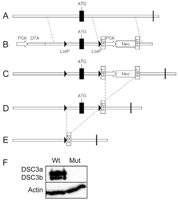
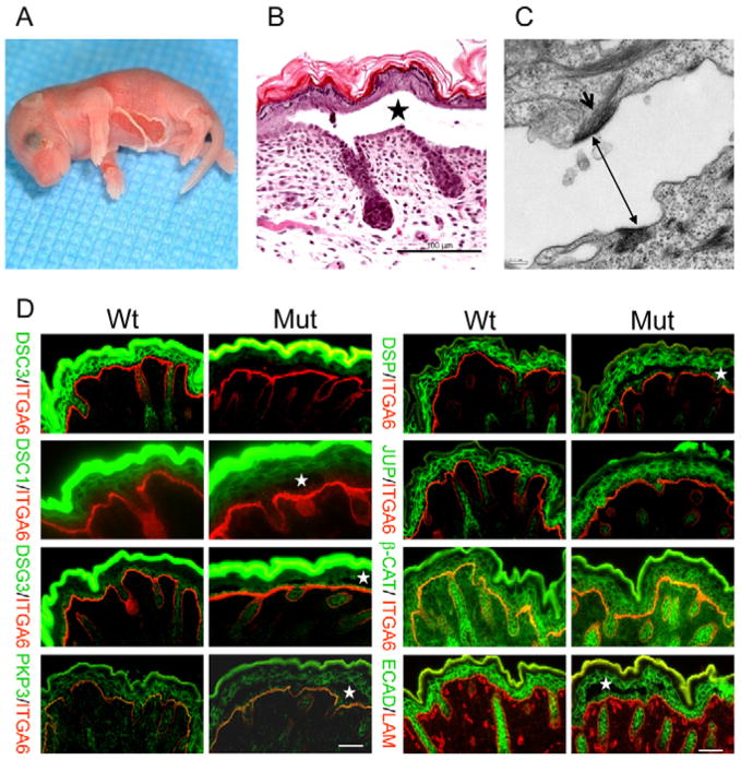
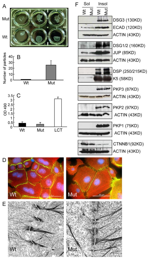
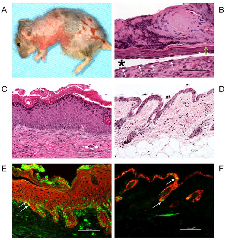
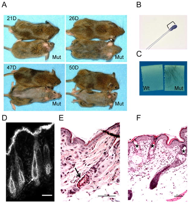
Desmocollin 3 (DSC3) belongs to a subfamily of cadherins and is a major component of desmosomes in keratinocytes of stratified epithelia, such as the epidermis. Based on its amino acid sequence homology to classical cadherins, such as E-cadherin, it has been postulated that DSC3 functions as a cell-adhesion molecule. To test this hypothesis, we assessed the function of DSC3 in the development and maintenance of stratified epithelia, in particular the epidermis and hair follicles. Using a conditional null allele, we show that loss of Dsc3 function in the epidermis causes impaired cell-cell adhesion, leading to intra-epidermal blistering and telogen hair loss. Furthermore, the lesions in Dsc3-null skin resemble those observed in individuals with pemphigus vulgaris (PV), indicating that impaired Dsc3 function could be a potential cause of PV-like inherited or acquired skin blistering diseases.


Figures

Fig. 1
Fig. 1
Schematic representation of the targeting strategy used to generate Dsc3fl (Dsc3tm2Ko) mice. (A) Exon 1 (ATG) of the Dsc3 gene was targeted with the vector shown in B. In the targeting construct, loxP sites were inserted in the promoter and intron 1. A neomycin minigene (PGK-Neo), flanked by FRT sites, was also inserted into intron 1. (C) The neo cassette was removed from recombinant ES cell clones via transient expression of FLPe recombinase, leaving a single FRT site in intron 1. (D) The resulting Dsc3fl/+ ES cells were used to generate mutant mice. (E) Cre-mediated recombination in bigenic mice (Dsc3fl/fl/K14-Cre) will lead to the deletion of exon 1, as well as part of the Dsc3 promoter and intron 1. (F) Newborn wild-type keratinocytes (Wt) synthesize both DSC3a and DSC3b, whereas Dsc3fl/fl/K14-Cre keratinocytes (Mut) do not express these proteins. The western blot shown was probed with a DSC3 antibody, stripped and re-probed with a β-actin antibody (loading control). Vertical bars indicate exons.
Fig. 2
Fig. 2
Characterization of newborn epidermis in mutant mice. (A) Newborn pup with severe ventral skin blistering. (B) Histology of an intra-epidermal blister in newborn mutant mice. Acantholysis is present immediately above the basal cell layer (star indicates blister cavity; bar, 100 μm). (C) Electron micrograph of a blister. Two half desmosomes are present in the plasma membranes of the acantholytic cells (double-headed arrow). The arrow indicates intermediate filaments that are attached to the half desmosome (bar, 0.2 μm). (D) Immunofluorescence microscopy using newborn skin of mutant mice (Mut) and wild-type (Wt) controls. The antibodies used are indicated. White stars indicate blister cavities. Junctional proteins are similarly distributed in both genotypes. ITGA6 (α6 integrin) or laminin staining (red) demarcates the basement membrane zone. The strong green fluorescence (DSC1, DSC3, DSG3 staining) is due to unspecific binding of the secondary antibody to the stratum corneum. Bars, 50 μm.
Fig. 3
Fig. 3
Weak cell adhesion between mutant keratinocytes. (A–C) Newborn mutant (Mut) and wild-type control (Wt) keratinocytes were grown in vitro to confluency, and then cultured in high Ca2+ medium for 24 hours to induce desmosome formation. The cell sheets were then lifted from the plate and exposed to mechanical stress (Huen et al., 2002). (A) Mutant cell sheets showed increased susceptibility to stress. Each well represents cells from a single pup, i.e. three mutant and three wild-type pups were used. (B) Quantitative evaluation of particles generated in the experiment shown above (average number of particles; n=3; error bar, standard deviation). (C) LDH release into the culture medium of stressed cell sheets. Mutant samples did not show increased cell lysis (LCT, lysis control; wild-type cells were treated with a detergent to release LDH; positive control). (D) Immunofluorescence staining of wild-type and mutant keratinocytes with antibodies against DSP (green) and K14 (red) (DAPI staining of the nucleus, blue). Bars, 50 μm. (E) Electron micrograph of desmosomes formed in newborn wild-type and mutant keratinocytes that were cultured in high Ca2+ medium for 24 hours. Morphologically normal desmosomes were formed in mutant keratinocytes. Bar, 0.5 μm. (F) Western blot analysis of newborn keratinocytes cultured as described in A. Detergent-insoluble (junction associated) and detergent-soluble (non-junctional) protein fractions were isolated as described (Cheng et al., 2004) and probed with the antibodies listed. β-Actin or keratin 5 (K5) were used as loading controls.
Fig. 4
Fig. 4
Skin phenotype of adult Dsc3fl/fl/K14-Cre mice. (A) A 160-day-old mutant mouse showing severe skin lesions, including a partial loss of the epidermis and hair follicles. (B,C) Histological analysis of the mouse shown in A. (B) Massive inflammation, scarring and re-epithelialization in an area where the epidermis was completely lost. A thin tongue of keratinocytes is migrating underneath the scab in an attempt to re-epithelialize the wound bed (the tongue is marked by a green double-headed arrow). The cells of this epithelial tongue undergo acantholysis (loss of cell–cell adhesion leading to blistering), a phenotype that might be the cause of wound healing defect and persistent skin blistering in older mice. Arrows indicate basal keratinocytes that have lost their adhesion to suprabasal cells, but that are still attached to the basement membrane. Owing to the severity of this phenotype, we now do not maintain these mice beyond 3 months of age. Star indicates blister cavity. (C) Hyperplastic area of mutant epidermis. (D) Age-matched normal skin of a wild-type mouse. The epidermis is only approximately two cell layers thick. (E) Immunofluorescence staining of hyperplastic mutant epidermis with K14 (red) and Ki67 (green) antibodies. The number of proliferating Ki67-positive nuclei (arrows) is dramatically increased compared with the wild-type control shown in F. (F) Age-matched wild-type epidermis stained with K14 (red) and Ki67 (green). Only a few keratinocytes in the interfollicular epidermis are Ki67 positive (arrows). Bars, 100 μm.
Fig. 5
Fig. 5
Hair-loss phenotype in Dsc3fl/fl/K14-Cre mice. (A) Beginning around the time of weaning, mutant mice developed hair loss that started at the head and proceeded towards the tail. (B,C) Tape-stripping experiments indicated telogen hair loss in mutant (Mut) but not wild-type (Wt) mice. A single cell layer of epithelial cells (stained blue with a histological dye in B) was still attached to the telogen hair club of loosely anchored mutant hair (bracket in B). (D). Staining of wild-type telogen skin with a DSC3 antibody. The two epithelial cell layers surrounding the club (arrow) express DSC3. (E) Acantholysis (arrow) between the two cell layers anchoring the telogen club hair in mutant skin. (F) Cyst formation (stars) in mutant skin resulting from telogen hair loss. Bars, 50 μm.

References

    1. Amagai M, Ahmed AR, Kitajima Y, Bystryn JC, Milner Y, Gniadecki R, Hertl M, Pincelli C, Kurzen H, Fridkis-Hareli M, et al. Are desmoglein autoantibodies essential for the immunopathogenesis of pemphigus vulgaris, or just “witnesses of disease”? Exp Dermatol. 2006;15:815–831. - PubMed
    1. Bolling MC, Mekkes JR, Goldschmidt WF, van Noesel CJ, Jonkman MF, Pas HH. Acquired palmoplantar keratoderma and immunobullous disease associated with antibodies to desmocollin 3. Br J Dermatol. 2007;157:168–173. - PubMed
    1. Bonne S, Gilbert B, Hatzfeld M, Chen X, Green KJ, van Roy F. Defining desmosomal plakophilin-3 interactions. J Cell Biol. 2003;161:403–416. - PMC - PubMed
    1. Chen J, Cheng X, Merched-Sauvage M, Caulin C, Roop DR, Koch PJ. An unexpected role for keratin 10 end domains in susceptibility to skin cancer. J Cell Sci. 2006;119:5067–5076. - PubMed
    1. Cheng X, Mihindukulasuriya K, Den Z, Kowalczyk AP, Calkins CC, Ishiko A, Shimizu A, Koch PJ. Assessment of splice variant-specific functions of desmocollin 1 in the skin. Mol Cell Biol. 2004;24:154–163. - PMC - PubMed

Publication types