. 2008 Aug 12;105(32):11224-9.
doi: 10.1073/pnas.0800620105. Epub 2008 Aug 5.

Promoter ChIP-chip analysis in mouse testis reveals Y chromosome occupancy by HSF2

Affiliations

Promoter ChIP-chip analysis in mouse testis reveals Y chromosome occupancy by HSF2

Malin Akerfelt et al. Proc Natl Acad Sci U S A. .

Abstract

The mammalian Y chromosome is essential for spermatogenesis, which is characterized by sperm cell differentiation and chromatin condensation for acquisition of correct shape of the sperm. Deletions of the male-specific region of the mouse Y chromosome long arm (MSYq), harboring multiple copies of a few genes, lead to sperm head defects and impaired fertility. Using chromatin immunoprecipitation on promoter microarray (ChIP-chip) on mouse testis, we found a striking in vivo MSYq occupancy by heat shock factor 2 (HSF2), a transcription factor involved in spermatogenesis. HSF2 was also found to regulate the transcription of MSYq resident genes, whose transcriptional regulation has been unknown. Importantly, disruption of Hsf2 caused a similar phenotype as the 2/3 deletion of MSYq, i.e., altered expression of the multicopy genes and increased mild sperm head abnormalities. Consequently, aberrant levels of chromatin packing proteins and more frequent DNA fragmentation were detected, implying that HSF2 is required for correct chromatin organization in the sperm. Our findings define a physiological role for HSF2 in the regulation of MSYq resident genes and the quality of sperm.


Conflict of interest statement

The authors declare no conflict of interest.

Figures

Fig. 1.
Fig. 1.
In vivo HSF2 binding to novel target genes in testis. (A) Visualization of the HSF2-binding profile on six selected target promoters, using the SignalMap software (NimbleGen Systems): Ssty2, Sly, Slx, Speer4a, Hsc70, and Ftmt. The localization of 15 probes per promoter is indicated as bars above the promoters, determining the HSF2 enrichment. One representative promoter is displayed for the multicopy genes. A putative heat shock element (HSE) is indicated below each promoter. Asterisk indicates key nucleotides required for HSF binding; arrows indicate primers used in the ChIP assay. Log2, log2 ratio of HSF2 enrichment, indicated as difference in positions of the bar in the log2 scale; Chr., chromosome; +1, transcription start site (note that the genes are transcribed in different directions). (B) Verification of the ChIP-chip screen using a standard ChIP assay of WT testis extracts. Analysis of HSF2-binding (HSF2) on the six selected promoters, in addition to three nontarget promoters for HSF2: Hsp25.1, Fyn, and Spata 2. (C) Target genes are occupied by HSF2 only in testis. ChIP analysis of HSF2-binding (HSF2) on six target promoters in WT testis, brain, muscle, and kidney. Nonspecific antibody (NS) was used as a negative control, and acetylated histone 4 antibody (AcH4) was used as an indicator of transcriptionally active promoters. Input represents 1% of the total material used in the ChIP assay.
Fig. 2.
Fig. 2.
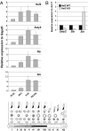
HSF2 functions as a transcriptional regulator of multicopy gene expression in spermatogenesis. (A) RT-PCR analysis of Hsf2, Ssty2, Sly, and Slx expression in the indicated stages of WT seminiferous epithelial cycle was performed. Relative quantities of mRNA were normalized to Gapdh, which was evenly expressed throughout the seminiferous epithelial cycle. A schematic presentation of the 12 stages (I–XII) in the mouse seminiferous epithelial cycle is shown below. Each stage is defined by a specific collection of cell types, which are classified by the morphology of the developing spermatids (21). (B) RT-PCR analysis of gene expression in whole WT (Hsf2 WT) and Hsf2 knockout (Hsf2 KO) testes. Relative quantities of mRNA were normalized to Acrv1/SP-10. All PCRs were in duplicates using samples derived from at least three biological repeats. Error bars denote standard deviations (±SD). The relative expression was calculated from the Hsf2 WT sample, which was arbitrarily set to 1.
Fig. 3.
Fig. 3.
HSF2-deficient mice display a significant increase in sperm head abnormalities. (A) Analysis of hematoxylin-stained sperm smears from adult WT (Hsf2 WT) and Hsf2 knockout (Hsf2 KO) males. Representative examples of different morphology are shown in the main figure, and a blow-up in the inset. (Scale bar, 10 μm.) (B) The sperm heads were classified in three categories of normal, slightly abnormal, and grossly abnormal, as previously described (23). The number of sperm in each category, obtained from Hsf2 WT (n = 4) and Hsf2 KO (n = 4) male mice, was calculated in blind. Error bars denote standard deviations (±SD).
Fig. 4.
Fig. 4.
Altered chromatin packing protein levels and increased DNA fragmentation in Hsf2 knockout sperm. (A) Western blot analysis of transition protein 2 (TNP2), protamine 1 (PRM1), and protamine 2 (PRM2) levels in cauda epididymis isolated from adult WT (Hsf2 WT) and Hsf2 knockout (Hsf2 KO) mice. The blots are representative examples of four biological repeats. Equal loading was assessed by β-tubulin and α-actin. (B) Analysis of DNA damage by comet assay. After separation of DNA fragments by electrophoresis, the sperm DNA from adult Hsf2 WT and Hsf2 KO males was stained with SYBR green. Representative examples of comets are shown with white arrows in the figure. (Scale bar, 10 μm.) (C) The percentages of sperm positive for DNA damage after analysis by comet assay were calculated from Hsf2 WT (n = 3) and Hsf2 KO (n = 3) mice. Error bars denote standard deviations (±SD).
Fig. 5.
Fig. 5.
A schematic presentation of HSF2 occupancy on the male-specific Y chromosome long arm (MSYq). The majority of the Y chromosome genes are located on the short arm (Yp) and were not found as HSF2 targets, whereas the MSYq mostly contains heterochromatin and repetitive sequences. All well annotated Y-chromosomal genes included in the ChIP-chip array are indicated in the figure. Note that the chromosome length and the number of HSF2 molecules are only illustrative. PAR, pseudoautosomal region.

References

    1. Ellis PJ, Affara NA. Spermatogenesis and sex chromosome gene content: An evolutionary perspective. Hum Fertil (Cambridge) 2006;9:1–7. - PubMed
    1. Toure A, et al. Identification of novel Y chromosome encoded transcripts by testis transcriptome analysis of mice with deletions of the Y chromosome long arm. Genome Biol. 2005;6:R102. - PMC - PubMed
    1. Sarge KD, Park-Sarge O-K, Kirby JD, Mayo KE, Morimoto RI. Expression of heat shock factor 2 in mouse testis: Potential role as a regulator of heat-shock protein gene expression during spermatogenesis. Biol Reprod. 1994;50:1334–1343. - PubMed
    1. Alastalo T-P, et al. Stage-specific expression and cellular localization of the heat shock factor 2 isoforms in the rat seminiferous epithelium. Exp Cell Res. 1998;240:16–27. - PubMed
    1. Kallio M, et al. Brain abnormalities, defective meiotic chromosome synapsis and female subfertility in HSF2 null mice. EMBO J. 2002;21:2591–2601. - PMC - PubMed

Publication types

MeSH terms

Associated data