. 2008 Aug 19;99(4):563-8.
doi: 10.1038/sj.bjc.6604504.

Human papillomavirus 'reflex' testing as a screening method in cases of minor cytological abnormalities

Affiliations

Human papillomavirus 'reflex' testing as a screening method in cases of minor cytological abnormalities

M Fröberg et al. Br J Cancer. .

Abstract

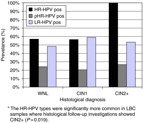
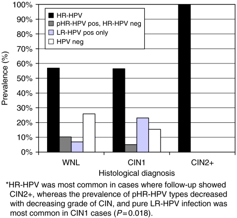
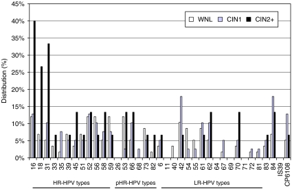
The aim was to evaluate human papillomavirus (HPV) 'reflex genotyping' in cases of minor cytological abnormalities detected in the gynaecological screening programme in Stockholm, Sweden. Liquid-based cytology samples showing minor cytological abnormalities were analysed using HPV genotyping (Linear Array, Roche diagnostics). Colposcopically directed cervical biopsies were obtained and the HPV test results were correlated with the histological results. In all, 63% (70/112) of the samples were high-risk (HR) HPV (HR-HPV) positive. A statistically significant correlation was found between high-grade cervical lesions and HR-HPV (P=0.019), among which HPV 16, 18, and 31 were the most important. The negative predictive value of HR-HPV detection for histologically confirmed high-grade lesions was 100%. An age limit for HPV reflex testing may be motivated in cases of low-grade squamous intraepithelial neoplasia (LSIL), because of high HR-HPV prevalence among younger women. By using HPV reflex genotyping, additional extensive workup can safely be avoided in about 50% of all cases of atypical squamous cells of undetermined significance (ASCUS) and LSIL among women 30 years. This screening strategy could potentially reduce the total abnormal cytology-reporting rate in the Swedish screening programme by about 1% and provide more accurately directed follow-up, guided by cytological appearance and HPV test results.


Figures

Figure 1
Figure 1
Prevalence of HPV risk categories related to histological diagnosis in 112 cases of minor cytological abnormalities. Single and multiple HPV infections are present within all risk categories. Since infection by HPV types of different risk categories overlap each other, the sum of the HR-, pHR-, and LR-HPV prevalences in each histological diagnosis group can exceed 100%. WNL (n=58)=within normal limits. CIN1 (n=39)=cervical intraepithelial neoplasia grade 1. CIN2+ (n=15): cervical intraepithelial neoplasia grade 2 or a more advanced lesion. HR-HPV pos (black bars)=samples positive for at least one HR-HPV type, with or without pHR- and/or LR-HPV coinfection. pHR-HPV pos (dark grey bars)=samples positive for at least one pHR-HPV type, with or without HR- and/or LR-HPV coinfection. LR-HPV pos (light grey bars)=samples positive for at least one LR-HPV type, with or without HR- and/or pHR-HPV coinfection.
Figure 2
Figure 2
Prevalence of HPV hierarchic risk categories related to histological diagnosis in 112 cases of minor cytological abnormalities. The HPV test results have been classified into risk categories according to the HPV type/types of the highest HPV risk category found in each sample. Using this description of the HPV infection pattern, there is no overlap between the different HPV risk categories. HR-HPV pos (black bars)=samples positive for at least one HR-HPV type, with or without pHR- and/or LR-HPV coinfection. pHR-HPV pos, HR-HPV neg (dark grey bars)=samples positive for at least one pHR-HPV type but negative for HR-HPV, with or without LR-HPV coinfection. LR-HPV pos only (light grey bars)= samples positive for at least one LR-HPV type, without HR- and pHR-HPV coinfection. HPV neg (white bars)=samples negative for all detectable HPV types.
Figure 3
Figure 3
Relative distribution of HR-, pHR-, and LR-HPV types in liquid-based cytology samples showing minor cytological abnormalities, related to histological diagnosis: WNL (n=58; light grey bars), CIN1 (n=39; dark grey bars), and CIN2+ (n=15; black bars). The corresponding figures are presented in detail in Supplementary Table 1.
Figure 4
Figure 4
Prevalence of single vs multiple HR-HPV infections in HR-HPV-positive cases related to histological diagnosis: WNL (n=33), CIN1 (n=22), and CIN2+ (n=15). Single HR-HPV infections (light grey bars). Multiple HR-HPV infections (black bars).

References

    1. Andersson S, Larson B, Hjerpe A, Silfverswärd C, Sällström J, Wilander E, Rylander E (2003) Adenocarcinoma of the uterine cervix: the presence of human papillomavirus and the method of detection. Acta Obstet Gynecol Scand 82: 960–965 - PubMed
    1. Andersson-Ellström A, Seidal T, Grannas M, Hagmar B (2000) The pap-smear history of women with invasive cervical squamous carcinoma. Acta Obstet Gynecol Scand 79: 221–226 - PubMed
    1. Andrae B, Kemetli L, Sparén P (2008) Screening-preventable cervical cancer risk: evidence for a nation-wide audit of cervical cancer in Sweden. J Nat Cancer Inst 100(9): 622–629 - PubMed
    1. Arbyn M, Sasieni P, Meijer CJ, Clavel C, Koliopoulos G, Dillner J (2006) Chapter 9: clinical applications of HPV testing: a summary of meta-analyses. Vaccine 24: 78–89 - PubMed
    1. Bergström R, Sparén P, Adami HO (1999) Trends in cancer of the cervix uteri in Sweden following cytological screening. Br J Cancer 81: 159–166 - PMC - PubMed

Publication types

MeSH terms