. 2008 Aug 6;3(8):e2869.
doi: 10.1371/journal.pone.0002869.

Protective immunity to Mycobacterium tuberculosis infection by chemokine and cytokine conditioned CFP-10 differentiated dendritic cells

Affiliations

Protective immunity to Mycobacterium tuberculosis infection by chemokine and cytokine conditioned CFP-10 differentiated dendritic cells

Nasir Salam et al. PLoS One. .

Abstract

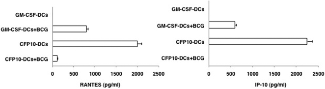
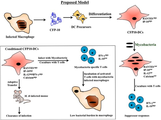
Background: Dendritic cells (DCs) play major roles in mediating immune responses to mycobacteria. A crucial aspect of this is the priming of T cells via chemokines and cytokines. In this study we investigated the roles of chemokines RANTES and IP-10 in regulating protective responses from Mycobacterium tuberculosis (M. tb) 10 kDa Culture Filtrate Protein-10 (CFP-10) differentiated DCs (CFP10-DCs).

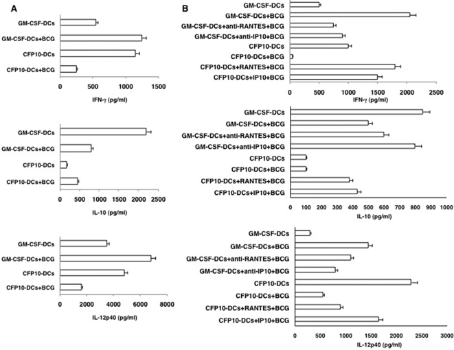
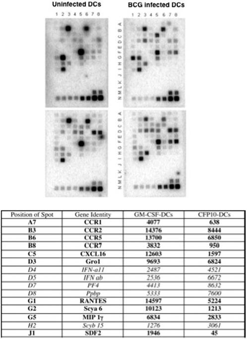
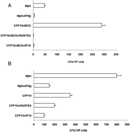
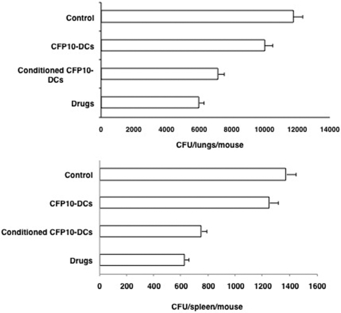
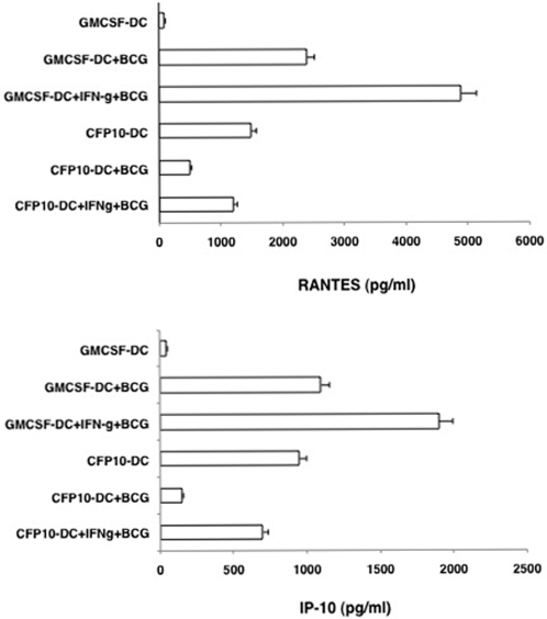
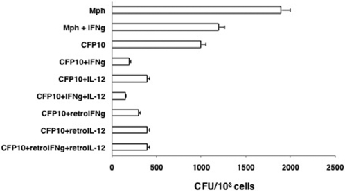
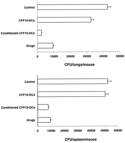
Methods and findings: Infection of CFP10-DCs with mycobacteria down-modulated RANTES and IP-10 levels. Pathway specific microarray analyses showed that in addition to RANTES and IP-10, mycobacteria infected CFP10-DCs showed reduced expression of many Th1 promoting chemokines and chemokine receptors. Importantly, T cells co-cultured with RANTES and IP-10 conditioned CFP10-DCs mediated killing of mycobacteria from infected macrophages. Similarly, T cells recruited by RANTES and IP-10 conditioned CFP10-DCs mediated significant killing of mycobacteria from infected macrophages. IFN-gamma treatment of CFP10-DCs restored RANTES and IP-10 levels and T cells activated by these DCs mediated significant killing of virulent M. tb inside macrophages. Adoptive transfer of either RANTES and IP-10 or IL-12 and IFN-gamma conditioned CFP10-DCs cleared an established M. tb infection in mice. The extent of clearance was similar to that obtained with drug treatment.

Conclusions: These results indicate that chemokine and cytokine secretion by DCs differentiated by M. tb antigens such as CFP-10 play major roles in regulating protective immune responses at sites of infection.


Conflict of interest statement

Competing Interests: The authors have declared that no competing interests exist.

Figures

Figure 1
Figure 1. CFP10-DCs downregulate RANTES and IP-10 levels following BCG infection.
GM-CSF-DCs or CFP10-DCs were infected with 1 MOI BCG for 24h. RANTES and IP-10 levels in culture supernatants were measured by ELISA. Data from one of five experiments is shown.
Figure 2
Figure 2. RANTES and IP-10 conditioned CFP10-DCs induce pro-inflammatory in vivo primary immune responses to BCG.
For Panel A GM-CSF-DCs or CFP10-DCs were infected in vitro with 1 MOI BCG for 24h. DCs were extensively washed to remove extracellular bacteria. 5×10⁶ DCs were adoptively transferred into naïve mice. Seven days later the inguinal lymph nodes were removed and 5×10⁶ cells/ml were cultured in 10% RPMI1640 medium with 10% FCS for 48h. Levels of indicated cytokines were measured in culture supernatants. For B, prior to infection with BCG, GM-CSF-DCs were either untreated or incubated with neutralizing monoclonal antibody to RANTES or IP-10 for 4h, while CFP10-DCs were either untreated or incubated with 25 ng/ml RANTES or IP-10 for 12h. DCs were extensively washed and then processed as in Panel A. Data from one of three experiments are shown.
Figure 3
Figure 3. RANTES and IP-10 elevate intracellular calcium in BCG stimulated CFP10-DCs.
FLUO-3-AM labeled GM-CSF-DCs (profile a–c) or CFP10-DCs (profile d–f) were stimulated with 1 MOI BCG and real-time increase in intracellular calcium was monitored over a period of 5 min. For profiles b and c GM-CSF-DCs were incubated with 5 μg/ml neutralizing monoclonal antibody to RANTES and IP-10, respectively, for 2h, prior to BCG stimulation. For profiles e and f, CFP10-DCs were incubated with 5 ng/ml recombinant RANTES and IP-10, respectively, for 12h, prior to BCG stimulation. DCs were extensively washed prior to flow cytometry. Data from one of three experiments are shown.
Figure 4
Figure 4. BCG infected CFP10-DCs show reduced expression of pro-inflammatory chemokines.
Total RNA was enriched from either GM-CSF-DCs (upper panels) or CFP10-DCs (lower panels) following infection with 1 MOI BCG (upper right and lower right panels) for 24h. 1 μg RNA was processed for microarray analyses using the GEArray Q Series mouse chemokine and receptor gene array from SuperArray strictly following the manufacturer's instructions. Rows A–I contains genes for various chemokines and their receptors while rows M and N contain house-keeping genes. Rows J–L represents negative controls. The Table below the figure depicts genes, their position and densitometric units of spots from blots of BCG infected GM-CSF-DCs and BCG infected CFP10-DCs. Data from one of two experiments are shown.
Figure 5
Figure 5. RANTES and IP-10 conditioned CFP10-DCs induce Th1 responses.
Either GM-CSF-DCs (GMCSF) or CFP10-DCs (CFP10) were infected with 1 MOI BCG for 24h and co-cultured for 48h with T cells enriched from BCG immunized mice. For some groups GM-CSF-DCs were incubated with 5 μg/ml neutralizing monoclonal antibody to RANTES and IP-10, respectively, for 2h, prior to BCG stimulation. CFP10-DCs were incubated with 5 ng/ml recombinant RANTES or IP-10 for 12h, prior to BCG stimulation. Data from one of three independent experiments are shown.
Figure 6
Figure 6. T cells co-cultured with or recruited by RANTES or IP-10 conditioned CFP10-DCs mediate clearance of BCG from macrophages.
For A, BCG infected CFP10-DCs (CFP10), conditioned or not with either 25 ng/ml RANTES or IP-10 were co-cultured for 48h with BCG primed T cells. From this, T cells were enriched and cultured with BCG infected macrophages (Mph) for 48h. A separate group wherein macrophages conditioned with 2 ng/ml IFN-γ (IFN-g) for 4h prior to infection with BCG was also included as a control. For B, T cells migrated into the lower chamber of a transwell apparatus in response to supernatants of BCG infected CFP-DCs, conditioned or not with 25 ng/ml RANTES or IP-10, were cultured with BCG infected macrophages (Mph) for 48h. A separate group wherein macrophages conditioned with 2 ng/ml IFN-γ (IFN-g) for 4h prior to infection with BCG was also included as a control. Cells from both Panels were lysed and plated in serial dilutions on 7H11 agar plates. CFU were counted 2–3 week later. Data are the mean±s.d. of three experiments. For A, P<0.002 (Mph vs CFP10+BCG+RANTES), P<0.004 (Mph vs CFP10+BCG+IP10). For B, P<0.02 (Mph vs CFP10+BCG+RANTES), P<0.01 (Mph vs CFP10+BCG+IP10).
Figure 7
Figure 7. RANTES and IP-10 conditioned CFP10-DCs mediate clearance of M. tb infection in vivo in mice.
Groups of mice were infected with 1×10⁶ M. tb H37Ra intravenously via the tail vein. Seven days post-infection, 10×106 unconditioned CFP10-DCs or CFP10-DCs conditioned with both RANTES and IP-10 together (25 ng/ml each) for 12h (conditioned CFP10-DCs) were injected into the tail vein of mice. A repeat injection was given 7 days following the first transfer. Seven days following the 2nd transfer, mice were sacrificed and lung and spleen homogenates were plated onto 7H11 agar plates in serial dilutions for CFU monitoring. In parallel, infected mice were injected with anti-TB drugs as given in Materials and Methods . Control represents mice infected with M. tb H37Ra followed by intravenous injection of PBS. Data are the mean±s.d. of three experiments. For lungs, P<0.02 (Control vs Conditioned CFP10-DCs), P<0.009 (CFP10-DCs vs conditioned CFP10-DCs). For spleen, P<0.006 (Control vs Conditioned CFP10-DCs), P<0.01 (CFP10-DCs vs conditioned CFP10-DCs).
Figure 8
Figure 8. Treatment with IFN-γ restores RANTES and IP-10 levels from BCG infected CFP10-DCs.
Either GM-CSF-DCs or CFP10-DCs were incubated with 2 ng/ml IFN-γ for 1h and washed. DCs were infected with 1 MOI BCG for 24h. Expression of RANTES and IP-10 was measured in culture supernatants. Data from one of three experiments are shown.
Figure 9
Figure 9. T cells co-cultured with IFN-γ or IL-12 conditioned CFP10-DCs mediate clearance of M. tb H37Rv from macrophages.
BCG infected CFP10-DCs (CFP10), conditioned or not with either 2 ng/ml IFN-γ (IFNg) or IL-12 or transformed with retroviruses expressing IFN-γ (retroIFNg) or IL-12p40 (retroIL-12), were co-cultured for 48h with BCG primed T cells. From this, T cells were enriched and cultured with M. tb H37Rv infected macrophages (Mph) for 48h. A separate group wherein macrophages conditioned with 2 ng/ml IFN-γ, prior to infection with M. tb H37Rv was also included as a control. Cells from all the groups were lysed and plated in serial dilutions on 7H11 agar plates for CFU monitoring 2–3 week later. Data are the mean±s.d. of three experiments. P<0.007 (Mph vs CFP10+IFNg), P<0.007 (Mph vs CFP10+IL-12); P<0.005 (Mph+IFNg vs CFP10+IFNg), P<0.005 (Mph+IFNg vs CFP10+IL-12); P<0.03 (CFP10 vs CFP10+IFNg), P<0.05 (CFP10 vs CFP10+IL-12).
Figure 10
Figure 10. IFN-γ or IL-12 conditioned CFP10-DCs mediate clearance of M. tb infection in vivo in mice.
Groups of mice were infected with 1×10⁶ M. tb H37Ra intravenously via the tail vein. Seven days post-infection, 10×106 unconditioned CFP10-DCs or CFP10-DCs treated with both IFN-γ and IL-12 together (2 ng/ml each) for 2h (conditioned CFP10-DCs) were injected into the tail vein of mice. A repeat injection with the same number of DCs was given 7 days following the first transfer. ‘Control’ represents mice infected with M. tb H37Ra followed by intravenous injection of PBS. Seven days following the 2nd transfer, mice were sacrificed and lung and spleen homogenates were plated onto 7H11 agar plates in serial dilutions for CFU monitoring. In parallel, infected mice were injected with anti-TB drugs as given in Materials and Methods . Data are the mean±s.d. of three experiments. For lungs, P<0.04 (Control vs Conditioned CFP10-DCs), P<0.03 (CFP10-DCs vs conditioned CFP10-DCs). For spleen, P<0.02 (Control vs Conditioned CFP10-DCs), P<0.02 (CFP10-DCs vs conditioned CFP10-DCs).
Figure 11
Figure 11. Conditioning CFP10-DCs with chemokines or cytokines induces protective responses.
Following infection by M. tb and their sequestration inside alveolar macrophages, antigens such as CFP-10 are secreted. CFP-10 induces the differentiation of DCs. CFP10-DCs secrete high levels of RANTES and IP-10. Following their interactions with mycobacteria CFP10-DCs reverse their phenotype and secrete low levels of RANTES, IP-10 and IL-12p40 and display poor calcium influx. This leads to increased survival of mycobacteria and the induction of suppressor T cell responses. Box, conditioning CFP10-DCs with either RANTES and IP-10 or IL-12 and IFN-γ induced pro-inflammatory T cell responses and T cells activated by these DCs mediate killing of M. tb inside infected macrophages. In addition, adoptive transfer of chemokine and cytokine conditioned CFP10-DCs into mice harboring an active M. tb infection results in clearance of infection that is commensurate with drug treatment.

Similar articles

Cited by

References

    1. World Health Organization. World Health Organization Fact Sheet, Geneva, Switzerland 2002.
    1. Steinman RM. Dendritic cells. In: William EPaul., editor. Fundamental Immunology. Lippincott-Raven Publishers, PA; 1999. pp. 547–573.
    1. Sousa CR. Dendritic cells as sensors of infection. Immunity. 2001;14:495–498. - PubMed
    1. Tian T, Woodworth J, Skold M, Behar SM. In vivo depletion of CD11c+ cells delays the CD4+ T cell response to Mycobacterium tuberculosis and exacerbates outcome of infection. J Immunol. 2005;175:3268–3272. - PubMed
    1. Banchereau J, Steinman RM. Dendritic cells and the control of immunity. Nature. 1998;392:245–52. - PubMed

Publication types

MeSH terms