

The yeast spore wall enables spores to survive passage through the digestive tract of Drosophila
- PMID: 18682732
- PMCID: PMC2478712
- DOI: 10.1371/journal.pone.0002873
The yeast spore wall enables spores to survive passage through the digestive tract of Drosophila
Abstract
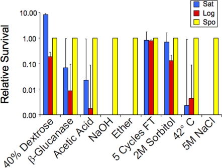
In nature, yeasts are subject to predation by flies of the genus Drosophila. In response to nutritional starvation Saccharomyces cerevisiae differentiates into a dormant cell type, termed a spore, which is resistant to many types of environmental stress. The stress resistance of the spore is due primarily to a spore wall that is more elaborate than the vegetative cell wall. We report here that S. cerevisiae spores survive passage through the gut of Drosophila melanogaster. Constituents of the spore wall that distinguish it from the vegetative cell wall are necessary for this resistance. Ascospores of the distantly related yeast Schizosaccharomyces pombe also display resistance to digestion by D. melanogaster. These results suggest that the primary function of the yeast ascospore is as a cell type specialized for dispersion by insect vectors.
Conflict of interest statement
Figures
Similar articles
-
Assay for Spore Wall Integrity Using a Yeast Predator.Cold Spring Harb Protoc. 2016 Aug 1;2016(8). doi: 10.1101/pdb.prot085258. Cold Spring Harb Protoc. 2016. PMID: 27480715
-
CRR1, a gene encoding a putative transglycosidase, is required for proper spore wall assembly in Saccharomyces cerevisiae.Microbiology (Reading). 2004 Oct;150(Pt 10):3269-80. doi: 10.1099/mic.0.27314-0. Microbiology (Reading). 2004. PMID: 15470107
-
The beta-1,3-glucanosyltransferase gas4p is essential for ascospore wall maturation and spore viability in Schizosaccharomyces pombe.Mol Microbiol. 2008 Jun;68(5):1283-99. doi: 10.1111/j.1365-2958.2008.06233.x. Epub 2008 Apr 8. Mol Microbiol. 2008. PMID: 18410286
-
Sporulation in the budding yeast Saccharomyces cerevisiae.Genetics. 2011 Nov;189(3):737-65. doi: 10.1534/genetics.111.127126. Genetics. 2011. PMID: 22084423 Free PMC article. Review.
-
Molecular and Biophysical Perspectives on Dormancy Breaking: Lessons from Yeast Spore.Biomolecules. 2025 May 11;15(5):701. doi: 10.3390/biom15050701. Biomolecules. 2025. PMID: 40427594 Free PMC article. Review.
Cited by
-
Reduction of Bacteria in Relation to Feeding Regimes When Treating Aquaculture Waste in Fly Larvae Composting.Front Microbiol. 2020 Jul 16;11:1616. doi: 10.3389/fmicb.2020.01616. eCollection 2020. Front Microbiol. 2020. PMID: 32765458 Free PMC article.
-
Assessment of fungal spores and spore-like diversity in environmental samples by targeted lysis.BMC Microbiol. 2023 Mar 14;23(1):68. doi: 10.1186/s12866-023-02809-w. BMC Microbiol. 2023. PMID: 36918804 Free PMC article.
-
Lipid droplet dynamics during Schizosaccharomyces pombe sporulation and their role in spore survival.Biol Open. 2017 Feb 15;6(2):217-222. doi: 10.1242/bio.022384. Biol Open. 2017. PMID: 28011631 Free PMC article.
-
Ingestion of genetically modified yeast symbiont reduces fitness of an insect pest via RNA interference.Sci Rep. 2016 Mar 2;6:22587. doi: 10.1038/srep22587. Sci Rep. 2016. PMID: 26931800 Free PMC article.
-
Microsatellite analysis of genetic diversity among clinical and nonclinical Saccharomyces cerevisiae isolates suggests heterozygote advantage in clinical environments.Mol Ecol. 2009 Jul;18(13):2779-86. doi: 10.1111/j.1365-294X.2009.04234.x. Epub 2009 May 20. Mol Ecol. 2009. PMID: 19457175 Free PMC article.
References
-
- Smits GJ, van den Ende H, Klis FM. Differential regulation of cell wall biogenesis during growth and development in yeast. Microbiology. 2001;147:781–794. - PubMed
-
- Kreger-Van Rij NJW. Electron microscopy of germinating ascospores of Saccharomyces cerevisiae. Arch Microbiol. 1978;117:73–77. - PubMed
-
- Briza P, Ellinger A, Winkler G, Breitenbach M. Chemical composition of the yeast ascospore wall. The second outer layer consists of chitosan. J Biol Chem. 1988;263:11569–11574. - PubMed
-
- Briza P, Winkler G, Kalchhauser H, Breitenbach M. Dityrosine is a prominent component of the yeast ascospore wall. A proof of its structure. J Biol Chem. 1986;261:4288–4294. - PubMed
Publication types
MeSH terms
Grants and funding
LinkOut - more resources
Full Text Sources
Other Literature Sources
Molecular Biology Databases
