. 2008 Aug 6;3(8):e2894.
doi: 10.1371/journal.pone.0002894.

Protein crystals in Adenovirus type 5-infected cells: requirements for intranuclear crystallogenesis, structural and functional analysis

Affiliations

Protein crystals in Adenovirus type 5-infected cells: requirements for intranuclear crystallogenesis, structural and functional analysis

Laure Franqueville et al. PLoS One. .

Abstract

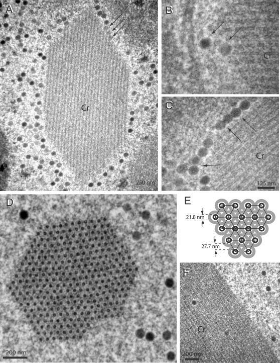
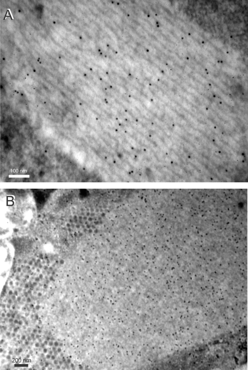
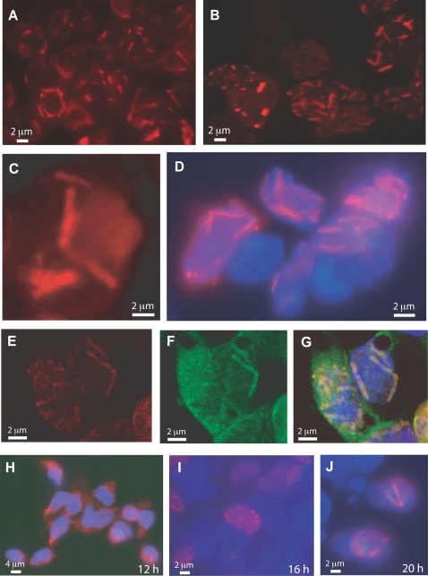
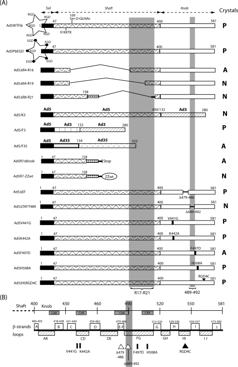
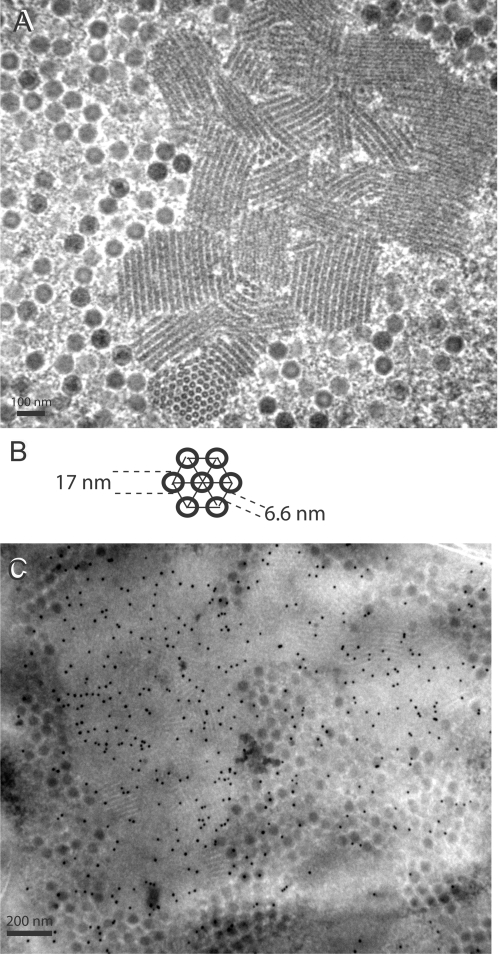
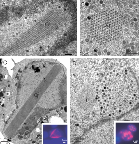
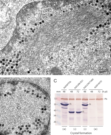
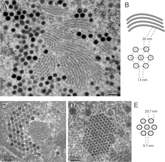
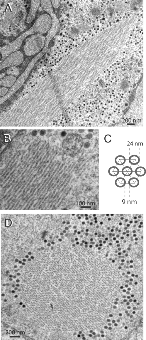
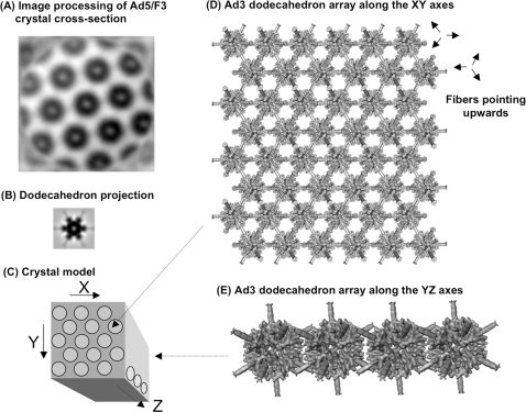
Intranuclear crystalline inclusions have been observed in the nucleus of epithelial cells infected with Adenovirus serotype 5 (Ad5) at late steps of the virus life cycle. Using immuno-electron microscopy and confocal microscopy of cells infected with various Ad5 recombinants modified in their penton base or fiber domains, we found that these inclusions represented crystals of penton capsomers, the heteromeric capsid protein formed of penton base and fiber subunits. The occurrence of protein crystals within the nucleus of infected cells required the integrity of the fiber knob and part of the shaft domain. In the knob domain, the region overlapping residues 489-492 in the FG loop was found to be essential for crystal formation. In the shaft, a large deletion of repeats 4 to 16 had no detrimental effect on crystal inclusions, whereas deletion of repeats 8 to 21 abolished crystal formation without altering the level of fiber protein expression. This suggested a crucial role of the five penultimate repeats in the crystallisation process. Chimeric pentons made of Ad5 penton base and fiber domains from different serotypes were analyzed with respect to crystal formation. No crystal was found when fiber consisted of shaft (S) from Ad5 and knob (K) from Ad3 (heterotypic S5-K3 fiber), but occurred with homotypic S3K3 fiber. However, less regular crystals were observed with homotypic S35-K35 fiber. TB5, a monoclonal antibody directed against the Ad5 fiber knob was found by immunofluorescence microscopy to react with high efficiency with the intranuclear protein crystals in situ. Data obtained with Ad fiber mutants indicated that the absence of crystalline inclusions correlated with a lower infectivity and/or lower yields of virus progeny, suggesting that the protein crystals might be involved in virion assembly. Thus, we propose that TB5 staining of Ad-infected 293 cells can be used as a prognostic assay for the viability and productivity of fiber-modified Ad5 vectors.


Conflict of interest statement

Competing Interests: The authors have declared that no competing interests exist.

Figures

Figure 1
Figure 1. EM analysis of Ad5WTFib-infected 293 cells at 48 h pi.
(A–C). Portions of cell nucleoplasm showing crystalline inclusions of viral proteins (Cr) seen in longitudinal sections. Note the presence of virions in close contact with crystal elements. Arrows on top of panel (A) indicate virions which seem to emerge from the edge of the crystal. Arrows on panel (B) and (C) point to filaments connecting virions to the crystal. (D), Portion of cell nucleoplasm showing a viral protein crystalline inclusion seen in cross section. (E), Model of the crystal lattice viewed in cross section, with its main parameters, diameter of the tubular unit (TD = 21.8) and inter-tubular distance (IT = 27.7), indicated in nm. In (F), a longitudinally sectioned crystal showed adenovirions included within the crystal lattice. Epon-embedded specimens.
Figure 2
Figure 2. EM and immuno-EM analysis of Ad5WTFib-infected 293 cells at 48 h pi.
Sections of metacrylate-embedded specimens were reacted with rabbit anti-fiber (A), or anti-penton base antibody (b), followed by 10-nm colloidal gold-labeled anti-rabbit IgG goat antibody. Shown are portions of nucleoplasm with protein crystalline inclusions. In panel (A), the longitudinal section of the protein crystal shows gold grains disposed along the lines of the tubular structures. In (B), crystalline inclusions of virus particles and viral protein are seen in close contact with each other, a phenomenon which is called epitaxy. Note that fixation and staining of metacrylate-embedded specimens was deliberately weak, in order not to interfere with the immunogold labeling.
Figure 3
Figure 3. IF microscopy of Ad-infected 293 cells at 48 h pi.
(A–D) and (H–J). Conventional IF microscopy using anti-fiber monoclonal antibody TB5 and secondary Alexa Fluor®633-labeled anti-mouse IgG. (A), Ad5WTFib; (B), Ad5PbEGD; (C), Ad5H508A; (D), Ad5(HI)RGD4C (with DAPI counterstaining). (E–G), Confocal analysis of Ad5ßGalWTFib-infected 293 cells with TB5. (E), Sample reacted with TB5 Alexa Fluor®633-labeled anti-mouse IgG; (F), sample reacted with anti-penton base antibody and secondary FITC-labeled anti-rabbit IgG; (G), image overlay with DAPI nuclear staining. (H–J), Kinetics of appearance of intranuclear crystals in Ad-infected cells. 293 cells infected with Ad5WTFib were harvested at different times pi and reacted with TB5 and Alexa Fluor®633-labeled anti-mouse IgG, with DAPI counterstaining. (H), 12 h pi; (I), 16 h pi; (J), 20 h pi. Note that IF pictures are presented at low magnification to show large field of view including several crystal-containing cells (A, B ; H–J), or at higher magnification to show the mass and number of crystals within a single nucleus (C–G).
Figure 4
Figure 4. (A), Schematic representation of penton base and fiber mutant constructs in Ad5 vectors.
The acronyms of the recombinants are indicated on the left side of the figure. The penton base and its RGD loops is represented by a five pointed star. The different structural domains and ligands of the fibers are shown by various symbols, as indicated in the figure. In Ad5ΔR8-21 and Ad5R7ΔKnob fibers, the extrinsic trimerization motif is represented by a stippled bar. On the right side of the figure, ‘Crystals’ indicates the occurrence of nuclear protein crystals : P, positive for crystals; N, negative; A, altered in crystal lattice arrangement. The shaded areas represent the regions of the fiber knob and shaft domains which are crucial for crystal formation. (B), Schematic representation of the Ad5 fiber knob domain and mutation positions. The end of the shaft domain is represented by a dotted line on the left side, and the knob domain by a solid line, with the amino acid numbering starting at residue 400. The β-strands regions are represented as open boxes, the flexible loops as hatched boxes, and the regions involved in CAR receptor binding as shaded boxes. The positions of mutations are indicated by solid bars for substitutions, by open triangles for deletions, and by a solid triangle for the RGD4C insertion.
Figure 5
Figure 5. EM (A), and immuno-EM (B) analyses of Ad5R7Δknob-infected 293 cells at 48 h pi.
In panel (A), an area of nucleoplasm shows small and twinned protein crystals seen in longitudinal, oblique and cross-sections. Virions are seen dispersed in the neighbourhood. Epon-embedded specimens. In panel (B), is shown a model of the crystal lattice derived from data obtained from as in (A). In panel (C), a section of metacrylate-embedded specimen was reacted with rabbit anti-penton base antibody followed by 10-nm colloidal gold-labeled anti-rabbit IgG goat antibody. Note the accumulation of gold-labeling in the protein crystalline inclusions. Some gold grains are also seen associated with virion clusters.
Figure 6
Figure 6. EM analysis of 293 cells infected with Ad fiber knob mutants at 48 h pi.
(A–C), Ad5ΔEF crystals (Cr) seen in longitudinal (A, C) and cross section (B); (C, inset), Ad5ΔEF-infected cells examined in IF microscopy after reaction with TB5 followed by Alexa Fluor®633-labeled anti-mouse IgG and DAPI counterstaining. (D), Ad5ΔTAYT489; (i), amorphous inclusion. (D, inset), Ad5ΔTAYT489-infected cells stained with TB5, as for the specimen shown in the inset of panel C.
Figure 7
Figure 7. Occurrence of intranuclear crystals and fiber protein expression in cells infected with Ad5 fiber mutants.
(A, B), EM analysis of Ad5F497D-infected cells at 48 h pi. Cr, crystal. (C), SDS-PAGE and Western blot analysis of whole lysates from cells infected respectively with : Ad5WTFib, Ad5ΔR8-R21, Ad5ΔTAYT489 and Ad5F497D, harvested at 48 or 72 h pi as indicated on top of the panel. Blots were reacted with anti-tail mAb 4D2.5 followed by phosphatase-labeled anti-mouse IgG antibody, then with anti-penton base rabbit antibody and peroxidase-labeled anti-rabbit IgG antibody. Fi, fiber polypeptide band; Pb, penton base. Symbols (+) and (−) at the bottom of the blot refer to Ad5 clones which were positive or negative for crystal formation, respectively.
Figure 8
Figure 8. EM analysis of 293 cells infected with (A, B) shaft-deleted recombinant Ad5ΔR4-R16, or (c–e) chimeric fiber recombinant Ad5/F3, at 48 h pi.
In panels (A) and (C, D), areas of nucleoplasm show small, twinned protein crystals seen in both longitudinal and cross-sections. Panels (B) and (E) present models of the two types of crystal lattice.
Figure 9
Figure 9. EM analysis of 293 cells infected with chimeric fiber recombinant Ad5/F35, at 48 h pi.
Protein crystals are shown in (A, B) longitudinal sections at low (A) and high (B) magnification, and (D) cross-section. (C), Model and parameters of the crystal lattice.
Figure 10
Figure 10. EM analysis of 293 cells infected with dual fiber-expressing recombinant Ad5/R7-ZZwt/E1:WTFib.
(A), Portion of nucleoplasm showing adenovirions clustering around a protein crystal (Cr) viewed in cross-section. (B), Another area of nucleoplasm showing Ad particles packed into clusters. Note that several particles have an electron-luscent centre (arrows).
Figure 11
Figure 11. Analysis of the Ad5/R7-ZZwt/E1:WTFib progeny.
(A–C), Hypothetical modes of intranuclear assembly for Ad5/R7-ZZwt/E1:WTFib virions. (A), Mode I : single population of mosaic fiber virions. (B), Mode II : two separate populations of virions, each carrying homogenous fiber species, WT and R7-ZZwt fiber, respectively. (C), Mode III : two separate populations of WT fiber virions and mosaic fiber virions, respectively. (D, E), EM analysis of Ad5/R7-ZZwt/E1:WTFib virus progeny. (D), Virus population banding at a density (ρ) of 1.345 in CsCl isopycnic gradient. (E), Virus population banding at ρ = 1.30–1.31. White arrows point to gaps in viral capsids. (F), Fiber protein content of the virus population banding at the densities of 1.345 and 1.31, respectively. Virions were analyzed by SDS-PAGE and Western blotting using anti-tail mAb 4D2.5 followed by (35S)-labeled anti-mouse IgG antibody. An autoradiogram is shown. The band labeled with (*) at 48 kDa corresponds to the reaction of secondary anti-mouse IgG antibody with core protein V. (G), High resolution EM of Ad5/R7-ZZwt/E1:WTFib progeny virions banding at ρ = 1.345. Black arrows point to long shafted, WT fibers. Red arrow points to a short shafted fiber.
Figure 12
Figure 12. EM analysis of Ad5/R7-ZZwt/E1:WTFib virus particles after immunogold labeling.
Ad5/R7-ZZwt/E1:WTFib particles of 1.34 in density were incubated with 6-nm gold-tagged polyclonal IgG from goat, and examined under the EM after negative staining with ammonium molybdate. The short, knob-deleted fibers R7-ZZwt were visualized by 6-nm gold-tagged goat IgG binding to the tandem ZZwt motif (IgG-Fc ligand). Long shafted WT fibers with their terminal knob are indicated by arrows.
Figure 13
Figure 13. Model of the crystal lattice of Ad penton intranuclear inclusions.
(A), 2D averaging of the Ad5/F3 crystal cross-section was generated from the crystal cross-section shown in Fig. 8 d. A total of 40 overlapping fields were cut out using the X3d program , and averaged after cross-correlation. (B), 2D projection of an Ad3 penton dodecamer (dodecahedron) with its 12 fiber projections, shown at the same magnification as in (a). The 3D map of a dodecahedron with 12 fibers was filtered to 45 Å and reprojected along its 3-fold axis using SPIDER . (C), Schematic 3D representation of a portion of penton crystal, showing its three axes (arrows). (D), Dodecahedron array, presented along the XY axes. (E), Arrangement of the dodecahedron units along the YZ axes. The 3D isosurface representation shown in (D) and (E) was visualized using WEB .

Similar articles

Cited by

References

    1. Russell WC. Update on adenovirus and its vectors. J Gen Virol. 2000;81:2573–2604. - PubMed
    1. Hong JS, Engler JA. Domains required for assembly of adenovirus type 2 fiber trimers. J Virol. 1996;70:7071–7078. - PMC - PubMed
    1. Li J, Lad S, Yang G, Luo Y, Iacobelli-Martinez M, et al. Adenovirus fiber shaft contains a trimerization element that supports peptide fusion for targeted gene delivery. J Virol. 2006;80:12324–12331. - PMC - PubMed
    1. Novelli A, Boulanger P. Deletion analysis of functional domains in baculovirus-expressed adenovirus type 2 fiber. Virology. 1991;185:365–376. - PubMed
    1. Novelli A, Boulanger PA. Assembly of adenovirus type 2 fiber synthesized in cell-free translation system. J Biol Chem. 1991;266:9299–9303. - PubMed

Publication types