. 2008 Aug 5;6(8):e191.
doi: 10.1371/journal.pbio.0060191.

TSCOT+ thymic epithelial cell-mediated sensitive CD4 tolerance by direct presentation

Affiliations

TSCOT+ thymic epithelial cell-mediated sensitive CD4 tolerance by direct presentation

Sejin Ahn et al. PLoS Biol. .

Abstract

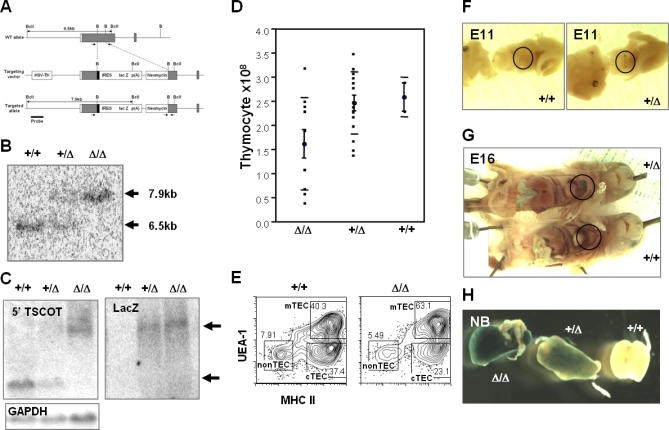
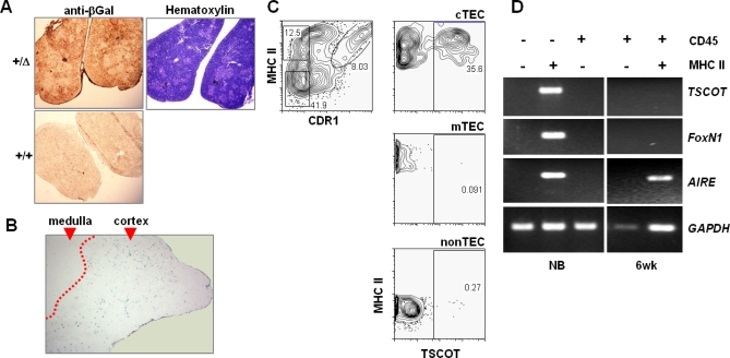
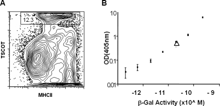
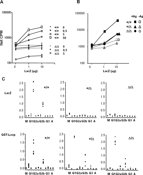
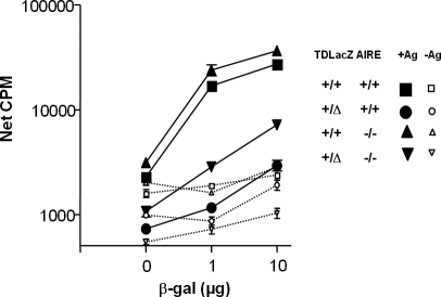
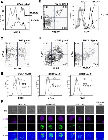
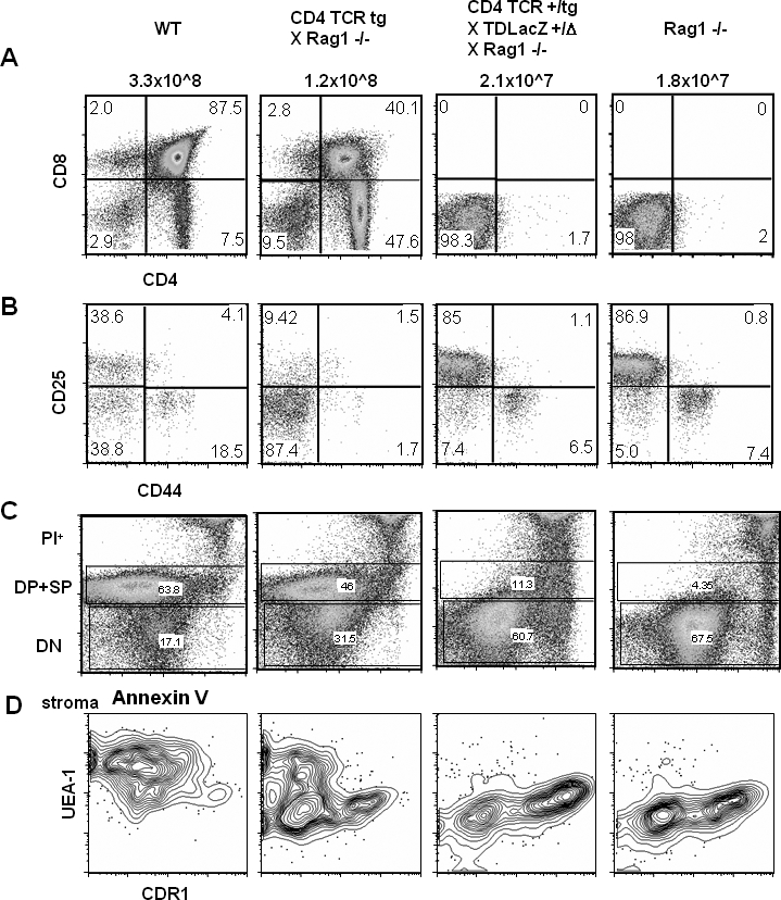
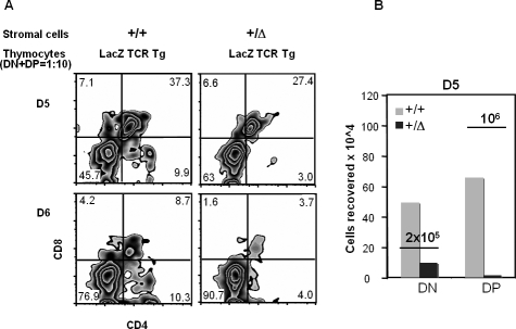
Although much effort has been directed at dissecting the mechanisms of central tolerance, the role of thymic stromal cells remains elusive. In order to further characterize this event, we developed a mouse model restricting LacZ to thymic stromal cotransporter (TSCOT)-expressing thymic stromal cells (TDLacZ). The thymus of this mouse contains approximately 4,300 TSCOT+ cells, each expressing several thousand molecules of the LacZ antigen. TSCOT+ cells express the cortical marker CDR1, CD40, CD80, CD54, and major histocompatibility complex class II (MHCII). When examining endogenous responses directed against LacZ, we observed significant tolerance. This was evidenced in a diverse T cell repertoire as measured by both a CD4 T cell proliferation assay and an antigen-specific antibody isotype analysis. This tolerance process was at least partially independent of Autoimmune Regulatory Element gene expression. When TDLacZ mice were crossed to a novel CD4 T cell receptor (TCR) transgenic reactive against LacZ (BgII), there was a complete deletion of double-positive thymocytes. Fetal thymic reaggregate culture of CD45- and UEA-depleted thymic stromal cells from TDLacZ and sorted TCR-bearing thymocytes excluded the possibility of cross presentation by thymic dendritic cells and medullary epithelial cells for the deletion. Overall, these results demonstrate that the introduction of a neoantigen into TSCOT-expressing cells can efficiently establish complete tolerance and suggest a possible application for the deletion of antigen-specific T cells by antigen introduction into TSCOT+ cells.


Conflict of interest statement

Competing interests. The authors have declared that no competing interests exist.

Figures

Figure 1
Figure 1. Targeting LacZ into the TSCOT Locus for Expression in Thymic Epithelium
(A) Schematic presentation of +/+ (top), the targeting construct (middle), and the targeted allele (bottom). The restriction sites BclI (BclI) and BamHI (B) and location of coding regions are shown. The probe (627 bp) used in the Southern blot is shown as a thick line under the targeted allele. PCR typing primer positions are shown as small arrows. (B) Southern blot of a BclI digest. The +/+ allele is 6.5 kb, and the targeted allele is 7.9 kb. (C) Northern blot for the TSCOT-LacZ fusion message. The probe (TSCOT, LacZ, and GAPDH control) is at the top left. (D) Total thymocyte yields from Δ/Δ, Δ/+, and +/+. Averages and standard deviations are shown. (E) Thymic stromal cell analyses of wild type and homozygotes. CD45 gates of 2-wk-old thymuses are shown for MHCII and UEA-1. The cTEC, mTEC, and nonTEC gates are indicated. (F and G) β-gal activity in the developing thymus (E11 and E16, respectively) of fetuses from Δ/+ and +/+ littermates. Only the targeted thymus was stained (within the black circles). Endogenous β-gal activity was detected in the intestines of both mice. (H) Newborn thymuses from Δ/Δ, Δ/+, and +/+ littermates show a gene dose-dependent expression.
Figure 2
Figure 2. LacZ and TSCOT-Expressing Cells in the Thymus
(A) Analyses of the thymus using an antibody against β-gal in newborns from a TDLacZ mouse heterozygote (Δ/+) and a wild type (+/+). Hematoxylin staining is shown on the right. (B) β-gal activity of the thymus from 8-wk-old homozygous knock-in mouse (Δ/Δ) at 10× magnification. Cortical and medullary areas are indicated, and the boundary between LacZ stained and unstrained areas is artificially marked as a dotted line for better visualization. (C) Flow cytometric analysis of TSCOT-expressing cells using the markers in total thymic CD45 stromal cells. The profiles of cortical marker CDR1 and medullary marker UEA-1. Defined cTEC, mTEC, and nonTEC are gated. Each gated population is shown as TSCOT and UEA-1 levels on the right. Fraction of TSCOT+ cells are shown in percentages. (D) Message expression of TSCOT, FoxN1, AIRE, and GAPDH by sorted thymic compartments according to CD45/MHCII status using RT-PCR (30 cycles). The cells were isolated either from newborns or 6-wk-old thymuses, and the cell surface markers used for sorting are shown on top.
Figure 3
Figure 3. Estimation of the Number of LacZ Molecules Expressed in the TSCOT-Expressing Cells
(A) Flow cytometric analysis of the percentage of TSCOT-expressing cells in the thymus preparation. (B) Standard curve of β-gal enzyme activity using purified LacZ protein. The purified LacZ enzyme from the commercial source was measured by the weight, and the stock enzyme solution was diluted serially to generate the curve. The open triangle indicates the measured value for a protein extract generated from 30 TDLacZ homozygous (Δ/Δ) thymuses.
Figure 4
Figure 4. CD4+ Tolerance to LacZ in the TDLacZ Mouse
(A) CD4+ T cells of mice immunized with 0, 0.5, 5, or 50 μg of LacZ, were stimulated with 0, 1, or 10 μg/ml of LacZ. The responses of LacZ immunized mice in solid lines with open symbols, the adjuvant immunized mice in dotted lines with closed symbols. Rectangles indicate +/+, and ovals Δ/Δ. (B) The T cells response of +/+ (rectangles), Δ/+ (triangles), and Δ/Δ (ovals) immunization with (solid lines and closed symbols) or without (dotted lines and open symbols) 25 μg of LacZ. There were no significant differences in the recovery of cell numbers from the immunized mice of any genotypes. (C) Antibody isotype profiles of +/+, +/Δ, and Δ/Δ mice immunized with LacZ (upper panels) or the GST-TSCOT-Loop (lower panels). The OD reading of individual serum is shown as a single symbol. Open symbols are for preimmune serum, and filled symbols are for immunized serum. IgM (M), IgG1 (G1) IgG2a (G2a), IgG2b (G2b), IgG3 (G3), and IgA (A) levels are shown.
Figure 5
Figure 5. AIRE Is Not Necessary for Tolerance to Antigens Expressed by TSCOT+ TECs
Proliferation assays in response to β-gal for lymph node CD4+ T cells from LacZ-immunized (10 μg) wild-type (rectangles), TDLacZ (ovals), or AIRE-deficient (triangles), or from AIRE-deficient × TDLacZ (inverted triangles) mice. The responses of the LacZ-immunized mice (+Ag) are shown as solid lines with closed symbols, while those of the adjuvant-only immunized mice (−Ag) are shown as dotted lines with open symbols.
Figure 6
Figure 6. Costimulatory Molecules and Other Surface Marker Profiling of TSCOT-Expressing Cells
(A) MHCII profiles of TSCOT+ cells (solid line) and TSCOT cells (dotted line). (B) CD40 profile of CD45 gate on the left, CD40. Histograms of TSCOT+, TSCOT(mTEC+cTEC), and CD45+ are indicated on the right. Background histogram of live gate is shown but not indicated. (C) CD54 profile of CD45 gate. (D) CD80 profile of CD45 gate and MHCII hi gate. (E) Histogram of CD80 levels in mTEC (UEA-1+CDR1), TSCOT+cTEC(CDR1+LacZ+), TSCOTcTEC (CDR1+LacZ). Specific CD80 stain (solid line) and background (dotted line) of the same gates are shown. Percentage of positive cells in each gates, MFI of negative and positive gates are indicated. (F) CD80 expression pattern of selected CDR1 and LacZ stained TDLacZ stromal cells. DIC, CDR1, LacZ, and CD80 staining patterns are shown.
Figure 7
Figure 7. Anti-LacZ–Specific TCR-Bearing Transgenic Thymocytes Are Deleted at the DN Stage in the Presence of TSCOT-LacZ Expression
(A) CD4 and CD8 thymocyte profiles. The names of each of the four mouse strains used and their total thymocyte yields are shown above the plots. The percentages of the single and double staining subsets are shown in the four quadrants. (B) CD25 and CD44 profiles of the DN thymocytes with percentages in the quadrants. (C) Annexin V staining of all cells stained with CD4, CD8, and PI shown in FL3 channel. PI+ cells are at the top, DP and SP cells in the middle, and DN cells in the bottom. (D) The UEA-1 and CDR1 profiles of the CD45 stromal cell compartment from the four different mouse lines. The genotypes of each mice are verified for the loci of Rag1, TDL, and TCR α and β chains by PCR
Figure 8
Figure 8. DC and mTEC Are Not Necessary for the Deletion of Transgenic Thymocytes
Sorted anti-LacZ TCR thymocytes were reaggregated with UEA-1–depleted CD45 stromal cells from dGuo-treated fetal thymuses. (A) Flow cytometric profiles of reaggregate cultures initiated with 3.8 × 10⁵ DN + DP (1:10 ratio) and 5 × 10⁵ purified thymic stroma at day 5 (top) or day 6 (bottom). The sources of stromal cells and thymocytes are indicated at the top of the graphs. CD4 and CD8 profiles are shown along with the percentage of cells in each of the compartments. Typically, cell recoveries of the DN and DP thymocyte mixtures cultured with TDLacZ heterozygote (+/Δ) stroma were 20%–30% of thymocyte mixtures cultured with wild-type (+/+) stroma. (B) A representative cell recovery when DN and DP cells were cultured separately with purified stroma from the wild type (+/+) versus the heterozygote (+/Δ ) mouse. Underlined numbers indicate the initial numbers of cells that were input.

Comment in

References

    1. Sprent J, Lo D, Gao EK, Ron Y. T cell selection in the thymus. Immunol Rev. 1988;101:173–190. - PubMed
    1. Ohashi PS. Negative selection and autoimmunity. Curr Opin Immunol. 2003;15:668–676. - PubMed
    1. Schwartz RH. T cell anergy. Annu Rev Immunol. 2003;21:305–334. - PubMed
    1. Arnold B, Schonrich G, Hammerling GJ. Multiple levels of peripheral tolerance. Immunol Today. 1993;14:12–14. - PubMed
    1. Coutinho A. Germ-line selection ensures embryonic autoreactivity and a positive discrimination of self mediated by supraclonal mechanisms. Semin Immunol. 2000;12:205–213. - PubMed

Publication types