Simulation of stent deployment in a realistic human coronary artery
- PMID: 18684321
- PMCID: PMC2525649
- DOI: 10.1186/1475-925X-7-23
Simulation of stent deployment in a realistic human coronary artery
Abstract
Background: The process of restenosis after a stenting procedure is related to local biomechanical environment. Arterial wall stresses caused by the interaction of the stent with the vascular wall and possibly stress induced stent strut fracture are two important parameters. The knowledge of these parameters after stent deployment in a patient derived 3D reconstruction of a diseased coronary artery might give insights in the understanding of the process of restenosis.
Methods: 3D reconstruction of a mildly stenosed coronary artery was carried out based on a combination of biplane angiography and intravascular ultrasound. Finite element method computations were performed to simulate the deployment of a stent inside the reconstructed coronary artery model at inflation pressure of 1.0 MPa. Strut thickness of the stent was varied to investigate stresses in the stent and the vessel wall.
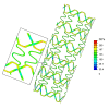
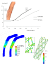
Results: Deformed configurations, pressure-lumen area relationship and stress distribution in the arterial wall and stent struts were studied. The simulations show how the stent pushes the arterial wall towards the outside allowing the expansion of the occluded artery. Higher stresses in the arterial wall are present behind the stent struts and in regions where the arterial wall was thin. Values of 200 MPa for the peak stresses in the stent strut were detected near the connecting parts between the stent struts, and they were only just below the fatigue stress. Decreasing strut thickness might reduce arterial damage without increasing stresses in the struts significantly.
Conclusion: The method presented in this paper can be used to predict stresses in the stent struts and the vessel wall, and thus evaluate whether a specific stent design is optimal for a specific patient.
Figures


References
-
- Serruys PW, de Jaegere P, Kiemeneij F, Macaya C, Rutsch W, Heyndrickx G, Emanuelsson H, Marco J, Legrand V, Materne P, Belardi J, Sigwart U, Colombo A, Goy JJ, Heuvel P van den, Delcan J, Morel MA. A comparison of balloon-expandable-stent implantation with balloon angioplasty in patients with coronary artery disease. Benestent Study Group. N Eng J Med. 1994;331:489–495. doi: 10.1056/NEJM199408253310801. - DOI - PubMed
-
- Rogers C, Edelman ER. Endovascular stent design dictates experimental restenosis and thrombosis. Circulation. 1995;91:2995–3001. - PubMed
-
- Duraiswamy N, Schoephoerster RT, Moreno MR, Moore JE. Stented artery flow patterns and their effects on the artery wall. Ann Rev Fluid Mech. 2007;39:357–382. doi: 10.1146/annurev.fluid.39.050905.110300. - DOI
Publication types
MeSH terms
LinkOut - more resources
Full Text Sources
Other Literature Sources

