. 2008 Sep 30;379(2):197-204.
doi: 10.1016/j.virol.2008.06.028. Epub 2008 Aug 6.

ICP34.5-dependent and -independent activities of salubrinal in herpes simplex virus-1 infected cells

Affiliations

ICP34.5-dependent and -independent activities of salubrinal in herpes simplex virus-1 infected cells

Kevin F Bryant et al. Virology. .

Abstract

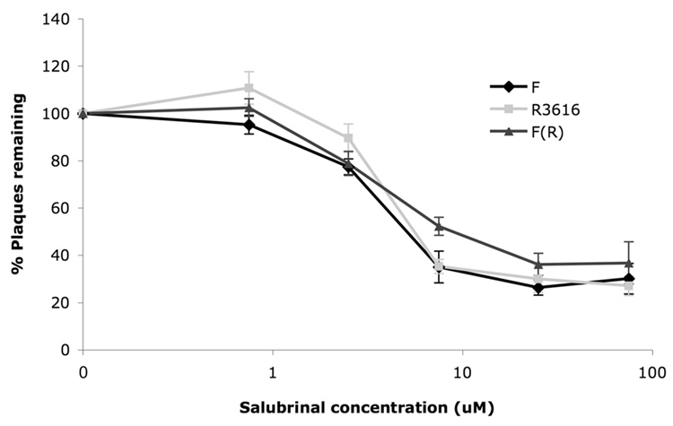
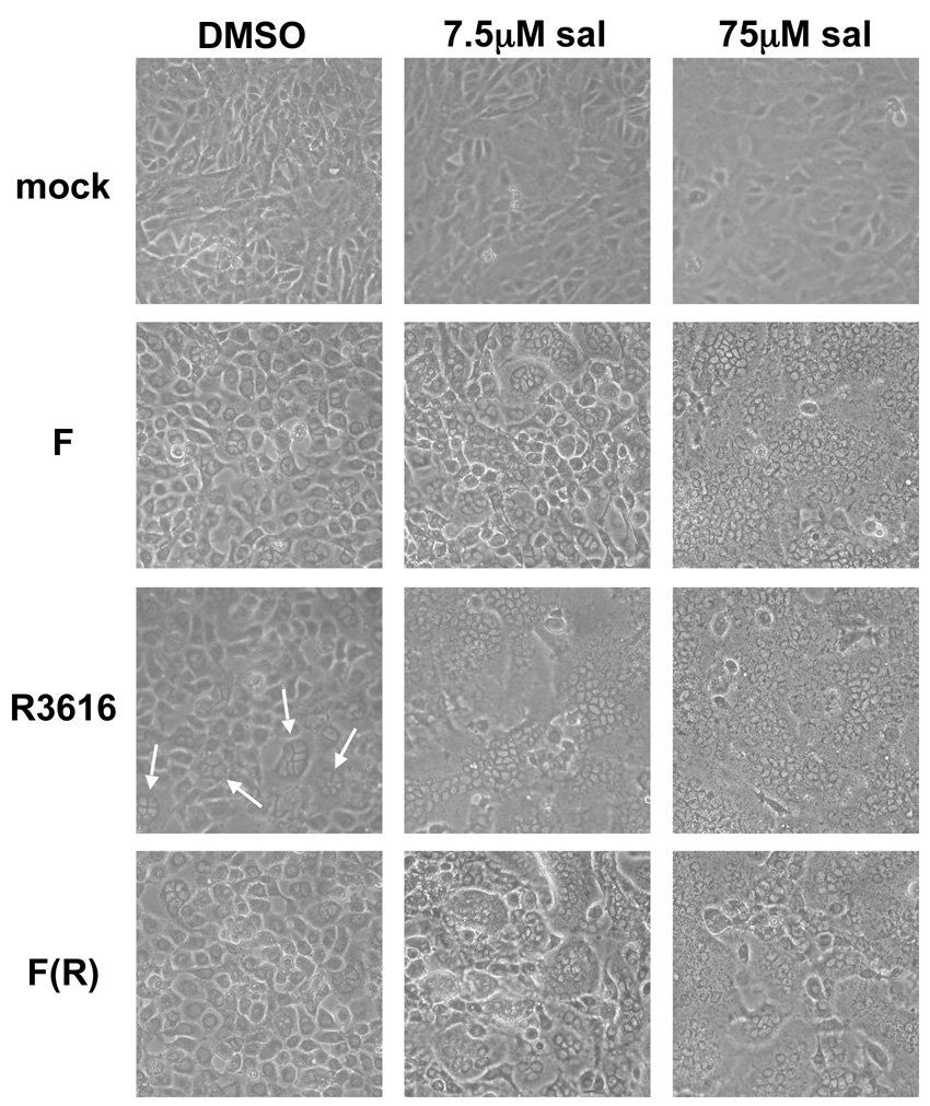
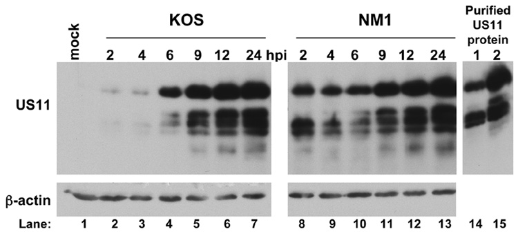
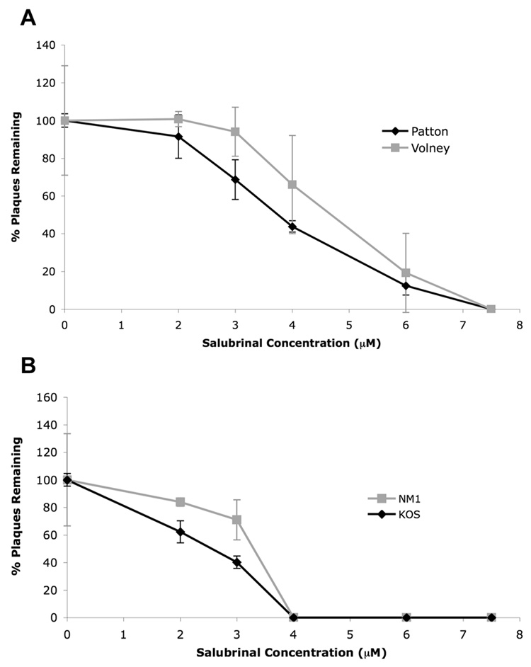
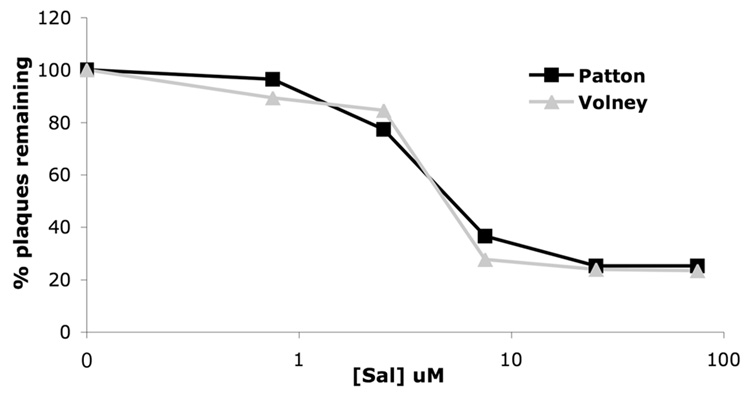
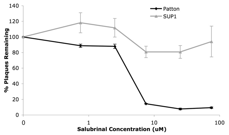
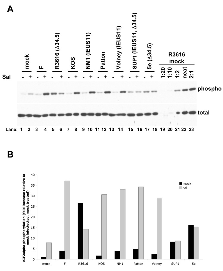
The small molecule salubrinal has antiviral activity against herpes simplex virus-1 (HSV-1) and inhibits dephosphorylation of eIF2 alpha mediated by the HSV-1 protein ICP34.5. We investigated whether salubrinal's activities in infected cells depend on ICP34.5. An ICP34.5 deletion mutant was as sensitive as wild type HSV-1 to salubrinal inhibition of plaque formation in Vero cells. However, salubrinal induced formation of syncytia in infected Vero cells, which was enhanced by ICP34.5 mutations. Expression of HSV-1 US11 with immediate early kinetics, which is known to suppress the effects of ICP34.5 mutations, resulted in slight resistance to salubrinal in murine embryonic fibroblasts, and substantial resistance in those cells when ICP34.5 was additionally mutated. ICP34.5 mutations, but not immediate early expression of US11, prevented salubrinal's ability to increase phosphorylation of eIF2 alpha during HSV-1 infection of Vero cells. Taken together, our data indicate that salubrinal has both ICP34.5-dependent and -independent activities in HSV-1 infected cells.


Figures

FIG. 1
FIG. 1. Antiviral activity of sal in plaque reduction assays
Vero cells were treated with either DMSO-containing vehicle or the indicated concentration of sal for 24 hours prior to infection with 150 pfu of HSV-1 strain F (black lines with diamonds), R3616 (light gray lines with squares), or F(R) (dark gray lines with triangles). Plaques were allowed to form for 2–3 days in the presence of the indicated concentration of sal and then were counted. Data are presented as percent of plaques remaining, relative to treatment with vehicle alone, at each sal concentration. Error bars represent standard errors of the mean (n=7 for strain F and R3616 and n=5 for F(R)). There was no significant difference (p values ranging between 0.072-0.97 by Student's t test) between F and R3616 at the various sal concentrations.
FIG. 2
FIG. 2. Sal induces syncytial HSV-1 infection
Vero cells were treated with either DMSO (left column) or the indicated concentration of sal (three right columns) for 24 hours prior to either mock infection (top row) or infection at a multiplicity of 3 with the indicated virus (bottom three rows). Infection was allowed to proceed for 18 hours in the presence of DMSO or the indicated concentration of sal and the cellular morphology was visualized by phase contrast light microscopy. Regions of small syncytia formation are indicated by arrows. Images were obtained by photographing with a Nikon Coolpix 4500 digital camera.
FIG. 3
FIG. 3. Western blot analysis of US11 expression
MEF cells were either mock infected (lane 1), infected with KOS at an MOI of 10 (lanes 2–7) or infected with NM1 at an MOI of 10 (lanes 8–13) and harvested at 2, 4, 6, 9, 12, or 24 hours post infection, as indicated above the top panel. Proteins in the cell lysates were resolved by SDS-polyacrylamide gel electrophoresis and transferred to a PVDF membrane. The membrane was cut and the bottom half was probed using an antibody against US11 while the membrane from the top half was probed with an antibody against β-actin to confirm equal loading among the samples. Purified recombinant US11 was also tested as a positive control for the US11 antibody and for migration of the immunoreactive species (lanes 14 and 15).
FIG. 4
FIG. 4. Slight resistance of the IE US11 viruses to sal in MEFs
MEF cells were pretreated with DMSO or varying concentrations of sal, as indicated, for 24 hours prior to infection with 150 pfu of (A) Patton (WT, black line with diamonds) and Volney (IE US11 virus, gray line with squares) or (B) KOS (WT, black line with diamonds) and NM1 (IE US11 virus, gray line with squares). After 3 days in the presence of sal, viral plaques were quantified and results show the percent of plaques remaining, relative to treatment with DMSO alone, at each sal concentration. Mean values from three separate experiments are shown (error bars represent standard errors of the mean).
FIG. 5
FIG. 5. Lack of resistance of the IE US11 virus to sal in Vero cells
Vero cells were treated with the indicated concentrations of sal for 24 hours prior to infection with 150 pfu of Patton (black line with squares), or Volney (gray line with triangles). Plaques were allowed to form for 2–3 days in the presence of the indicated concentration of sal and were then counted. Data are presented as the percentage of plaques remaining, relative to the DMSO treatment, at each sal concentration.
FIG. 6
FIG. 6. Resistance of SUP1 to sal in MEFs
MEF cells were pretreated with DMSO or varying concentrations of sal, as indicated, for 24 hours prior to infection with 150 pfu of Patton (WT, black line with diamonds) and SUP1 (IE US11, Δ34.5 virus, gray line with triangles). After 3 days in the presence of sal, viral plaques were quantified and results show the percent of plaques remaining, relative to DMSO treatment alone, at each sal concentration. Data shown are from a single experiment run in triplicate. Error bars represent standard errors of the mean.
FIG. 7
FIG. 7. Effects of salubrinal on eIF2α phosphorylation
(A) Western blot analysis of eIF2α phosphorylation. Vero cells were treated with either 75µM sal (+, lanes 2, 4, 6, 8, 10, 12, 14, 16, and 18) or DMSO-containing vehicle (−, lanes 1, 3, 5, 7, 9, 11, 13, 15, and 17) for 24 hours prior to either mock infection (lanes 1 and 2) or infection with HSV-1 strain F (lanes 3 and 4), the ICP34.5 mutant R3616 (lanes 5 and 6), KOS (lanes 7 and 8), the IE US11 mutant NM1 (lanes 9 and 10), Patton (lanes 11 and 12), the IE US11 mutant Volney (lanes 13 and 14), the IE US11, ICP34.5 double mutant SUP1 (lanes 15 and 16), or the ICP34.5 mutant 5e (lanes 17 and 18) at an MOI of 3 in the presence of DMSO or sal. At 14 hours post infection, lysates were prepared and resolved by SDS-PAGE. Samples were resolved on duplicate gels and were immunoblotted for either total eIF2α (bottom panel) or serine-51 phosphorylated eIF2α (top panel). (B) Quantification of the western blot data in (A). The dilution series at the right end of each panel (lanes 19−23) were used to generate a standard curve by determining the intensity of each band on a scanned film using Quantity One software (BioRad). The standard curve was then used to determine the relative concentration of phosphorylated or total eIF2α in each sample. The level of phosphorylated eIF2α was normalized to the level of total eIF2α in each sample. The results are presented as the fold increase in phosphorylation relative to the normalized level of phosphorylated eIF2α in the mock treated, mock infected sample.

References

    1. Boyce M, Bryant KF, Jousse C, Long K, Harding HP, Scheuner D, Kaufman RJ, Ma D, Coen DM, Ron D, Yuan J. A selective inhibitor of eIF2alpha dephosphorylation protects cells from ER stress. Science. 2005;307:935–939. - PubMed
    1. Boyce M, Yuan J. Cellular response to endoplasmic reticulum stress: a matter of life or death. Cell Death Differ. 2006;13:363–373. - PubMed
    1. Brown SM, MacLean AR, Aitken JD, Harland J. ICP34.5 influences herpes simplex virus type 1 maturation and egress from infected cells in vitro. J Gen Virol. 1994;75:3679–3686. - PubMed
    1. Bryant KF, Cox JC, Wang H, Hogle JM, Ellington AD, Coen DM. Binding of herpes simplex virus-1 US11 to specific RNA sequences. Nucleic Acids Res. 2005;33:6090–6100. - PMC - PubMed
    1. Cassady KA, Gross M. The herpes simplex virus type 1 US11 protein interacts with protein kinase R in infected cells and requires a 30-amino-acid sequence adjacent to a kinase substrate domain. J Virol. 2002;76:2029–2035. - PMC - PubMed

Publication types

MeSH terms