. 2009 Mar 13;284(11):7149-56.
doi: 10.1074/jbc.M801681200. Epub 2008 Aug 6.

Constitutively activated ALK2 and increased SMAD1/5 cooperatively induce bone morphogenetic protein signaling in fibrodysplasia ossificans progressiva

Affiliations

Constitutively activated ALK2 and increased SMAD1/5 cooperatively induce bone morphogenetic protein signaling in fibrodysplasia ossificans progressiva

Toru Fukuda et al. J Biol Chem. .

Abstract

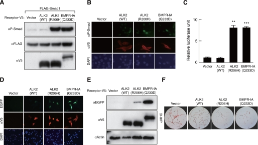
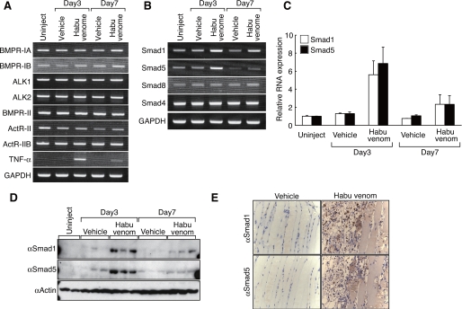
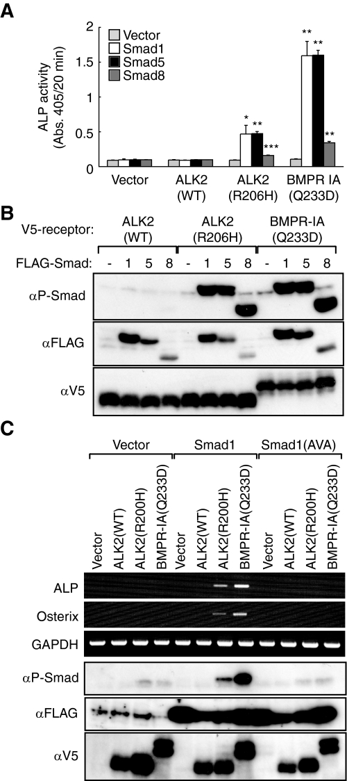
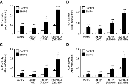
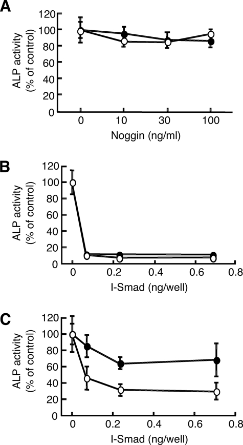
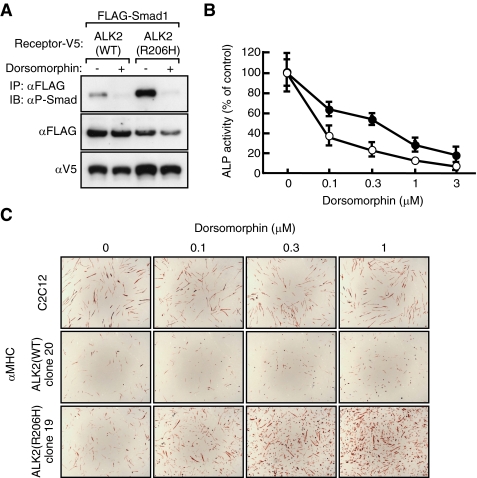
Fibrodysplasia ossificans progressiva (FOP) is a rare autosomal dominant disorder characterized by congenital malformation of the great toes and by progressive heterotopic bone formation in muscle tissue. Recently, a mutation involving a single amino acid substitution in a bone morphogenetic protein (BMP) type I receptor, ALK2, was identified in patients with FOP. We report here that the identical mutation, R206H, was observed in 19 Japanese patients with sporadic FOP. This mutant receptor, ALK2(R206H), activates BMP signaling without ligand binding. Moreover, expression of Smad1 and Smad5 was up-regulated in response to muscular injury. ALK2(R206H) with Smad1 or Smad5 induced osteoblastic differentiation that could be inhibited by Smad7 or dorsomorphin. Taken together, these findings suggest that the heterotopic bone formation in FOP may be induced by a constitutively activated BMP receptor signaling through Smad1 or Smad5. Gene transfer of Smad7 or inhibition of type I receptors with dorsomorphin may represent strategies for blocking the activity induced by ALK2(R206H) in FOP.


Figures

FIGURE 1.
FIGURE 1.
ALK2(R206H) acts as a constitutively activated BMP receptor. A, C2C12 cells were co-transfected with FLAG-tagged Smad1 and a V5-tagged wild-type ALK2 (WT), ALK2(R206H), or BMPR-IA(Q233D). Cell lysates were immunoblotted with anti-phospho-Smad1/5/8, anti-FLAG, or anti-V5 antibody. Constitutively active BMPR-IA(Q233D) was used as a positive control. B, C2C12 cells transfected with wild-type ALK2 or ALK2(R206H) were immunostained with anti-phospho-Smad1/5/8 or anti-V5 antibody and 4′,6-diamidino-2-phenylindole (DAPI). C, C2C12 cells were co-transfected with IdWT4F-luc reporter plasmid and wild-type ALK2, ALK2(R206H), or BMPR-IA(Q233D). Results are the means ± S.D. (n = 3). **, p < 0.01; ***, p < 0.001 compared with vector transfection. D and E, C2C12 cells were co-transfected with Id-EGFPd2 reporter plasmid and wild-type ALK2, ALK2(R206H), or BMPR-IA(Q233D). Levels of enhanced green fluorescent protein were determined by fluorescence microscopy (D) and immunoblotting (E). F, C3H10T1/2 cells co-transfected with a MyoD expression construct (24) and empty vector, wild-type ALK2, ALK2(R206H), or BMPR-IA(Q233D) were stained with anti-MHC antibody.
FIGURE 2.
FIGURE 2.
Increased Smad1 and Smad5 mRNA during muscular injury in vivo. A-C, mice were injected with vehicle (saline) or habu venom in femoral muscle, and total RNA was prepared after 3 or 7 days. Messenger RNA levels of BMP receptors (A) and Smads (B and C) were determined by RT-PCR (A and B) or real time quantitative PCR (C). Tumor necrosis factor-α (TNF-α) expression was examined to confirm inflammatory reaction. D, levels of Smad1 and Smad5 proteins in injured muscle in vivo were detected by immunoblotting at 3 and 7 days after injury. Two and three independent mice were analyzed in the control (uninjected) and vehicle and Habu venom-injected groups, respectively. E, localization of Smad1 and Smad5 in muscle tissues was determined by immunohistochemistry using specific antibodies to Smad1 and Smad5, respectively, on day 3 after injection. GAPDH, glyceraldehyde-3-phosphate dehydrogenase.
FIGURE 3.
FIGURE 3.
Cooperative effect of ALK2(R206H) with Smad1/5 in induction of osteoblastic differentiation. A and B, C2C12 cells were co-transfected with FLAG-tagged Smad1, Smad5, or Smad8 with V5-tagged wild-type ALK2(WT), ALK2(R206H), or BMPR-IA(Q233D). ALP activity (A) and levels of phosphorylation of Smads (B) were determined on day 3. Results are the means ± S.D. (n = 3). *, p < 0.05; **, p < 0.01; ***, p < 0.001 compared with vector transfection in each group. C, C2C12 cells were co-transfected with FLAG-tagged Smad1 or Smad1(AVA) and V5-tagged wild-type ALK2, ALK2(R206H), or BMPR-IA(Q233D). RT-PCR was performed to determine levels of expression of ALP and osterix mRNAs after 3 days. Levels of phosphorylated Smads and receptors were determined by immunoblotting using anti-phospho-Smad1/5/8, anti-FLAG, or anti-V5 antibody (lower panels). GAPDH, glyceraldehyde-3-phosphate dehydrogenase.
FIGURE 4.
FIGURE 4.
BMPs further stimulate ALP activity induced by ALK2(R206H) and Smad1 in C2C12 myoblasts. C2C12 cells co-transfected with Smad1 and wild-type (WT) ALK2, ALK2(R206H), or BMPR-IA(Q233D) were treated for 3 days with 300 ng/ml of BMP-2 (A) or 100 ng/ml of BMP-4 (B), BMP-6 (C), or BMP-7 (D), and ALP activity was then determined. Results are the mean ± S.D. (n = 3). *, p < 0.05; **, p < 0.01; ***, p < 0.001 compared with controls.
FIGURE 5.
FIGURE 5.
Inhibition of biological activities of ALK2(R206H) by Noggin and I-Smads. A, C2C12 cells co-transfected with Smad1 and ALK2(R206H) (closed circles) or BMPR-IA(Q233D) (open circles) were treated with Noggin. ALP activity was determined on day 3. B and C, C2C12 cells were co-transfected with BMPR-IA(Q233D) (B) or ALK2(R206H) (C) and Smad1, with increasing amounts of Smad6 (closed circles) or Smad7 (open circles). Total amounts of DNA transfected were adjusted equally with an empty vector. ALP activity was determined on day 3. Results are the mean ± S.D. (n = 3).
FIGURE 6.
FIGURE 6.
Dorsomorphin inhibits ALK2(R206H) activity. A, HEK293 cells were co-transfected with FLAG-tagged Smad1 and V5-tagged ALK2(R206H) or V5-BMPR-IA(Q233D) and then treated for 3 days with dorsomorphin at 3 μm. Levels of phosphorylated Smad1 were determined by immunoblotting (IB). IP, immunoprecipitation. B, C2C12 cells were co-transfected with Smad1 and ALK2(R206H) (closed circles) or BMPR-IA(Q233D) (open circles) and treated with graded concentrations of dorsomorphin. ALP activity was determined on day 3. C, parental C2C12 cells, C2C12-ALK2 (WT) clone 20, and C2C12-ALK2(R206H) clone 19 were treated with increasing concentrations of dorsomorphin in low serum medium and stained with anti-MHC antibody on day 3.

References

    1. Cohen, R. B., Hahn, G. V., Tabas, J. A., Peeper, J., Levitz, C. L., Sando, A., Sando, N., Zasloff, M., and Kaplan, F. S. (1993) J. Bone Jt. Surg. Am. 75 215-219 - PubMed
    1. Kaplan, F. S., McCluskey, W., Hahn, G., Tabas, J. A., Muenke, M., and Zasloff, M. A. (1993) J. Bone Jt. Surg. Am. 75 1214-1220 - PubMed
    1. Kaplan, F. S., Tabas, J. A., Gannon, F. H., Finkel, G., Hahn, G. V., and Zasloff, M. A. (1993) J. Bone Jt. Surg. Am. 75 220-230 - PubMed
    1. Kaplan, F. S., Shen, Q., Lounev, V., Seeman, P., Groppe, J., Katagiri, T., Pignolo, R. J., and Shore, E. M. (2008) J. Bone Miner. Metab. 26 521-530 - PMC - PubMed
    1. Kaplan, F. S., Glaser, D. L., Shore, E. M., Pignolo, R. J., Xu, M., Zhang, Y., Senitzer, D., Forman, S. J., and Emerson, S. G. (2007) J. Bone Jt. Surg. Am. 89 347-357 - PubMed

Publication types

MeSH terms

LinkOut - more resources