Review
. 2008 Aug 15;5(6):675-81.
doi: 10.1513/pats.200801-006AW.

Lung epithelial progenitor cells: lessons from development

Affiliations
Review

Lung epithelial progenitor cells: lessons from development

Emma L Rawlins. Proc Am Thorac Soc. .

Abstract

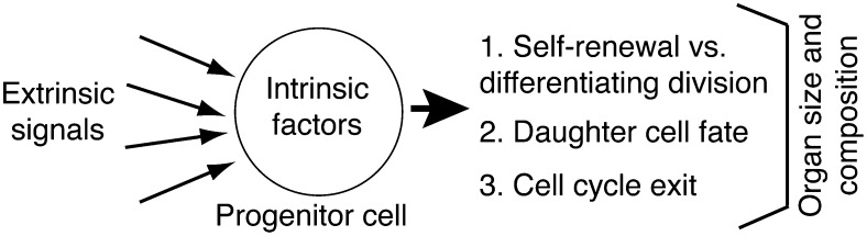
The current enthusiasm for stem cell research stems from the hope that damaged or diseased tissues may one day be repaired through the manipulation of endogenous or exogenous stem cells. The postnatal human respiratory system is highly accessible and provides unique opportunities for the application of such techniques. Several putative adult lung epithelial stem cells have been identified in the mouse model system. However, their in vivo capabilities to contribute to different lineages, and their control mechanisms, remain unclear. If stem cell-based therapies are to be successful in the lung, it is vitally important that we understand the normal behavior of adult lung stem cells, and how this is regulated. Lung embryonic progenitor cells are much better defined and characterized than their adult counterparts. Moreover, experiments on a variety of developing tissues are beginning to uncover general mechanisms by which embryonic progenitors influence final organ size and structure. This provides a framework for the study of lung embryonic progenitor cells, facilitating experimental design and interpretation. A similar approach to investigating adult lung stem cells could produce rapid advances in the field.


Figures

Figure 1.
Figure 1.
Aspects of tissue morphogenesis potentially controlled by regulation of progenitor cell behavior. Progenitor cells can control various aspects of tissue morphogenesis and hence influence organ size and structure. These include (1) the number of progenitor cell divisions and whether these are symmetric or asymmetric, (2) the final differentiated fate that daughter cells acquire, (3) whether daughter cells exit the cell cycle or continue to proliferate. Progenitor cells themselves are regulated both by extrinsic signaling and intrinsic factors determined by their developmental history and in turn signal back to their neighbors. These characteristics are common to both embryonic progenitors and adult stem cells.
Figure 2.
Figure 2.
A multipotent epithelial progenitor cell population is localized within the distal epithelial buds throughout lung development. (A) Confocal image of Embryonic Day 14.5 (E14.5) FoxJ1-GFP lung. E-cadherin (blue) labels the epithelium. Progenitor cells (Sox9+, red) are located at the budding tips of the epithelium. As the lung branches and cells exit the distal tips, they turn off Sox9 and other distal epithelium-specific genes, become committed to a specific lineage, and begin to differentiate. Differentiating cells (in this example, green fluorescent protein [GFP+] ciliated cells; green) are located proximally to the tips. Bar = 50 μm. (B, C) The conducting airways are laid down between E10 and E16 and the alveoli between E17 and E18. A multipotent progenitor population is located at the distal epithelial tips throughout lung development and therefore must transition from generating conducting airway cells to generating alveolar cells. There are several possible mechanisms by which this may occur. (B) Progenitors are multipotent throughout lung development and the fate of their progeny is completely dependent on extrinsic signaling. (C) There is only one population of progenitor cells, but its competence to give rise to different cell types changes over the course of development.
Figure 3.
Figure 3.
Neuroendocrine (NE) cell fate specification. During development, cells are believed to acquire a specific differentiated identity via a multistep process. (A) Expression of transcription factors and local signaling result in a cell becoming specified to differentiate along a particular lineage. For example, cells that express high levels of Ascl1 and do not respond to Notch signaling become the NE cell precursors. (B) The precursor expresses genes, often transcription factors, that commit its architecture to its final differentiated fate. High levels of ascl1, neuroD1, and gfi1 appear to be important at this stage of NE cell differentiation. (C) The final differentiated cell maintains a specific morphology and pattern of gene expression appropriate for its role in the tissue. At this stage the NE cells express high levels of calcitonin gene-related peptide (CGRP) and uchl1 (ubiquitin carboxy-terminal hydrolase 1, also known as PGP9.5).

References

    1. Noctor SC, Martinez-Cerdeno V, Ivic L, Kriegstein AR. Cortical neurons arise in symmetric and asymmetric division zones and migrate through specific phases. Nat Neurosci 2004;7:136–144. - PubMed
    1. Guillemot F. Spatial and temporal specification of neural fates by transcription factor codes. Development 2007;134:3771–3780. - PubMed
    1. Johansson KA, Dursun U, Jordan N, Gu G, Beermann F, Gradwohl G, Grapin-Botton A. Temporal control of neurogenin3 activity in pancreas progenitors reveals competence windows for the generation of different endocrine cell types. Dev Cell 2007;12:457–465. - PubMed
    1. Cardoso WV, Lu J. Regulation of early lung morphogenesis: questions, facts and controversies. Development 2006;133:1611–1624. - PubMed
    1. Okubo T, Knoepfler PS, Eisenman RN, Hogan BL. Nmyc plays an essential role during lung development as a dosage-sensitive regulator of progenitor cell proliferation and differentiation. Development 2005;132:1363–1374. - PubMed

Publication types

LinkOut - more resources