. 2008 Oct;82(20):10231-46.
doi: 10.1128/JVI.01890-07. Epub 2008 Aug 6.

Functions of Varicella-zoster virus ORF23 capsid protein in viral replication and the pathogenesis of skin infection

Affiliations

Functions of Varicella-zoster virus ORF23 capsid protein in viral replication and the pathogenesis of skin infection

Vaishali Chaudhuri et al. J Virol. 2008 Oct.

Abstract

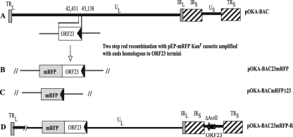
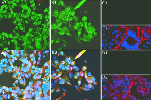
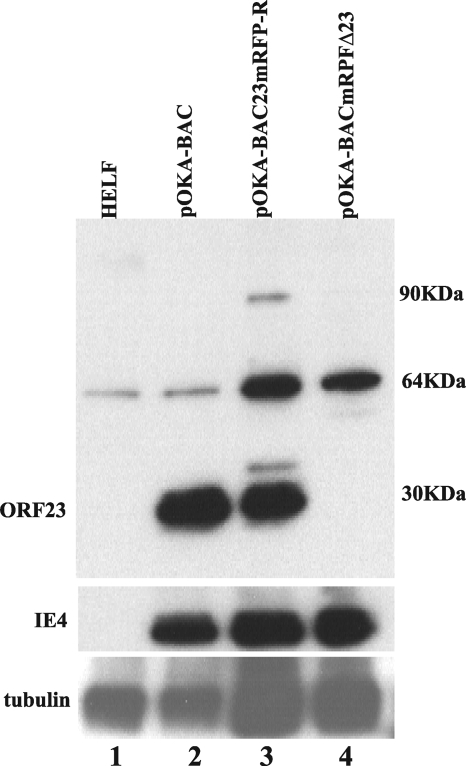
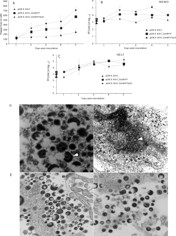
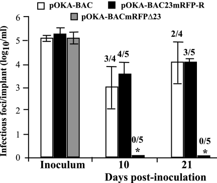
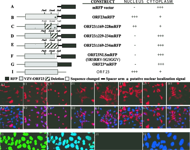
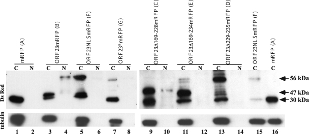
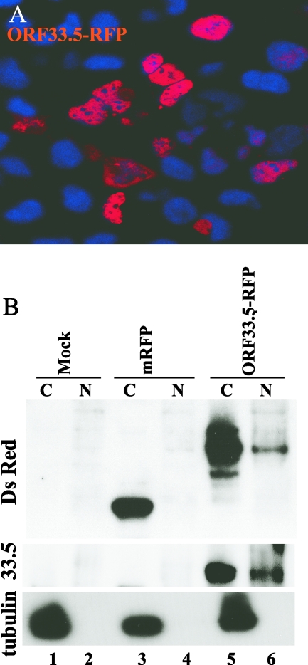
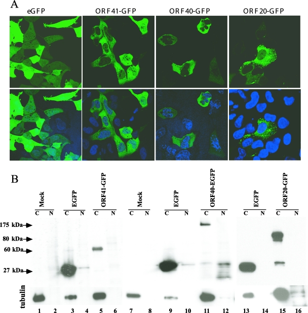
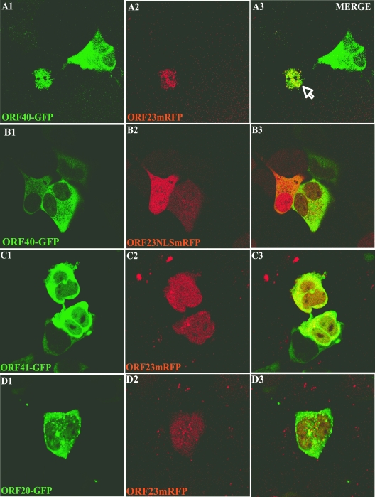
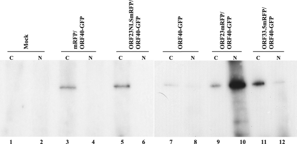
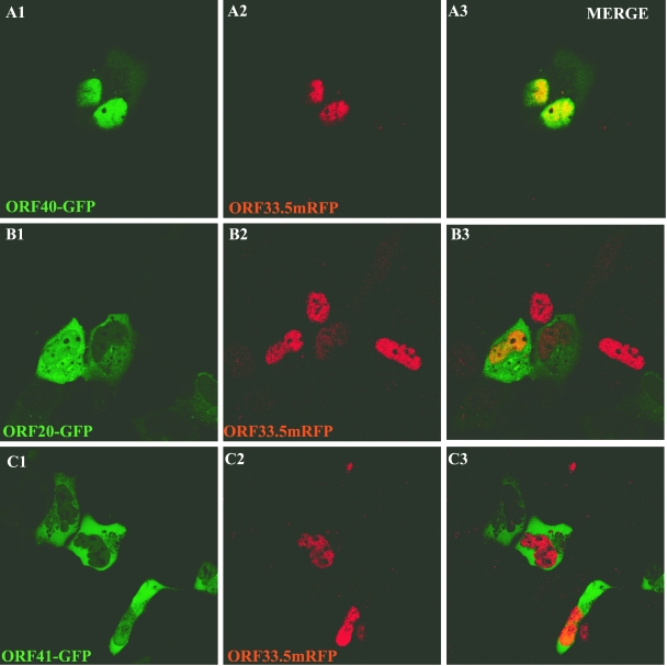
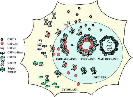
The assembly of herpesvirus capsids is a complex process involving interactions of multiple proteins in the cytoplasm and in the nucleus. Based on comparative genome analyses, varicella-zoster virus (VZV) open reading frame 23 (ORF23) encodes a conserved capsid protein, referred to as VP26 (UL35) in other alphaherpesviruses. Mutagenesis using a VZV bacterial artificial chromosome system showed that ORF23 was dispensable for replication in vitro. However, the absence of ORF23 disrupted capsid assembly in a melanoma cell line. Expression of ORF23 as a red fluorescent protein (RFP) fusion protein appeared to have a dominant negative effect on replication that was rescued by ORF23 expression from a nonnative site in the VZV genome. In contrast to its VP26 homolog, ORF23 has an intrinsic nuclear localization capacity that was mapped to an SRSRVV motif at residues 229 to 234 in the extreme C terminus of ORF23. In addition, coexpression with ORF23 resulted in nuclear import of the major capsid protein, ORF40. VZV ORF33.5 also translocated ORF40, which may provide a redundant mechanism in vitro but appears insufficient to overcome the dominant negative effect of the monomeric RFP-ORF23 (mRFP23) fusion protein. ORF23 was required for VZV infection of human skin xenografts, indicating that ORF33.5 does not compensate for lack of ORF23 in vivo. These observations suggest a model of VZV capsid assembly in which nuclear transport of the major capsid protein and associated proteins requires ORF23 during VZV replication in the human host. If so, ORF23 expression could be a target for a novel antiviral drug against VZV.


Figures

FIG. 1.
FIG. 1.
Schematic representation of ORF23 mutagenesis in pOKA-BAC. A. Location of ORF23 in the unique long region (UL) segment of the VZV genome; the terminal repeats (TR), unique short region (US), and internal repeats (IR) are indicated. The arrow shows the direction of ORF23 transcription. B. Insertion of mRFP (gray box) just before the ORF23 stop codon. C. Replacement of ORF23 with mRFP. D. ORF23 insertion at the ectopic AvrII site in the US (position 112,851) of pOKA-BAC23mRFP (dark box).
FIG. 2.
FIG. 2.
ORF23 protein expression in MeWo cells infected with pOKA-BAC, pOKA-BAC23mRFP-R, and pOKA-BACmRFPΔORF23. MeWo cells infected with pOKA-BAC (A1 and A2), pOKA-BAC23mRFP-R (B1 and B2), or pOKA-BACmRFPΔ23 (C1 and C2) were stained with rabbit anti-ORF23 IgG (1:2,000) and detected with fluorescein isothiocyanate-labeled goat anti-rabbit IgG (top panel) and merged with staining with human polyclonal anti-VZV IgG (1:1,000) detected with Texas red-labeled goat anti-human IgG and Hoechst nuclear stain (bottom panel). The preimmune rabbit IgG showed no reactivity with MeWo cells infected with OKA-BAC (D1 and D2).
FIG. 3.
FIG. 3.
ORF23 expression in HELF cells infected with pOKA-BAC, pOKA-BAC23mRFP-R, or pOKA-BACmRFPΔ23. Uninfected and infected HELF lysates were run on a 10% SDS-polyacrylamide gel and probed with anti-ORF23 (1:2,000). Lane 1, uninfected HELF; lane 2, pOKA-BAC; lane 3, pOKA-BAC23mRFP-R; lane 4, pOKA-BACmRFPΔ23. The blots were probed with anti-IE4 to show similar viral protein loading, which required adding more infected cell lysate for the ORF23 mutants (center panel). Anti-α-tubulin (1:16,000) was used to assess cell protein loading, which was higher, as expected, in lanes with lysates of ORF23 mutant-infected HELF (lower panel).
FIG. 4.
FIG. 4.
Plaque morphologies and growth kinetics of pOKA-BAC and ORF23 mutants in MeWo and HELF cells. (A) Plaque diameters were measured in MeWo cells infected with ∼1 × 10³ PFU of pOKA-BAC, pOKA-BAC23mRFP-R, and pOKA-BACmRFPΔ23 from days 1 through 5. The x axis indicates the days after inoculation when infected cell monolayers were stained with anti-VZV IgG, and the y axis indicates the size of the plaques in micrometers. (B and C) MeWo cells (B) and HELF (C) cells were inoculated with pOKA-BAC23mRFP-R and pOKA-BACmRFPΔ23 at ∼1 × 10³ PFU. Infectious virus yields were determined from days 1 to 5 after infection. Titers are expressed as means for triplicate wells at each time point. The x axes indicate the days after inoculation when infected cell monolayers were harvested, and the y axes indicate PFU determined by infectious focus assay. (D) Analysis of virion formation of ORF23 mutants and pOKA BAC in Mewo cells by TEM. Left panel: accumulation of typical capsids in the nucleus of pOKA-BAC-infected MeWo cells 48 h after inoculation. Magnification, ×35,000. Right panel: accumulation of empty spherical particles in the nucleus of pOKA-BACmRFPΔ23-infected MeWo cells; white arrows point to the empty spherical capsids. Magnification, ×35,000. (E) Analysis of virion formation of ORF23 mutants and pOKA BAC in HELF cells by TEM. Left panel: accumulation of typical capsids in the nucleus of pOKA-BAC-infected HELF cells 48 h after inoculation. Magnification, ×10,000. Right panel: accumulation of capsids in the nuclei of pOKA-BACmRFPΔ23-infected HELF cells. Magnification, ×10,000.
FIG. 5.
FIG. 5.
Replication of pOKA-BAC and ORF23 mutants in skin xenografts in SCIDhu mice. Skin xenografts were inoculated with pOKA-BAC, pOKA-BAC23mRFP-R, and pOKA-BACmRFPΔ23. Each bar represents the mean titer of infectious virus (error bars show standard deviations) recovered from four to five xenografts harvested at 10 and 21 days after inoculation. Inoculum titers are shown at the left. The asterisk indicates no viral growth in any implants. The number of xenografts per number inoculated from which infectious virus was recovered is shown above each bar. Mean titers were based on data from xenografts that yielded virus.
FIG. 6.
FIG. 6.
Analysis of ORF23 localization using transient-expression constructs. (Top) The schematic illustrates the mRFP vector (A), the ORF23mRFP fusion protein (B), truncations (C, D, and E), and point mutations (F) in the sequence encoding ORF23 fused to mRFP or ORF23 directly fused to mRFP without any linker (G), and ORF23 not fused to mRFP (I). The solid black box indicates the mRFP vector, and the gray shaded box indicates the ORF23 sequence. The * marks the location of the putative nuclear localizing signal, the hatched box indicates regions that were deleted, and the dark box indicates amino acid changes from the intact ORF23 protein. The ORF23 cassette is joined to the mRFP fusion protein with a 16-amino-acid spacer arm, indicated by the solid black line, which is part of the vector backbone. The names of the constructs and their subcellular localizations when expressed in AD293 cells are indicated to the right of each schematic. (Bottom panel) Detection of cellular localization of mRFP in AD293 cells transfected with ORF23 wild-type and mutant constructs and controls by direct fluorescence confocal microscopy at 32 h after transfection. The alphabetical notation in the panels (i.e., A1, B1, C1, D1, E1, F1, G1, and I1) correspond with each construct mentioned at the top, and the corresponding lower panel (A2, B2, C2, D2, E2, F2, G2, and I2) shows merged images of the same construct with nuclear Hoechst stain. (H1) Direct fluorescence images from HSV-1 VP26mRFP; (H2) same construct merged with Hoechst nuclear stain. (I1) AD293 cells transfected with pDS-ORF23, stained with rabbit anti-ORF23 IgG (1:2,000), was detected with fluorescein isothiocyanate-labeled goat anti-rabbit IgG; (I2) merged image with Hoechst nuclear stain. Control panels J1 and J2 depict background fluorescence in AD293 cells with anti-ORF23 IgG.
FIG. 7.
FIG. 7.
Immunoblot analysis of the intracellular localization of ORF23 and ORF23 mutant proteins. Control vector, ORF23, and ORF23 mutant constructs were transfected into AD293 cells, and the cell lysate was separated into cytoplasmic (C) and nuclear (N) fractions. Blots were probed with polyclonal dsRED antibody. ORF23mRFP expression yielded a 56-kDa band; mRFP protein expressed alone was 30 kDa. Blots were probed with anti-α-tubulin as a control for separation of cytoplasmic and nuclear fractions. Letters A to G in parentheses following the construct names correspond to the letters in the confocal images in Fig. 6. Lanes: 1 and 2, vector control; 3 and 4, ORF23-RFP; 5 and 6, ORF23NLSmRFP; 7 and 8, ORF23*RFP; 9 and 10, ORF23Δ169-228mRFP; 11 and 12, ORF23Δ169-234mRFP; 13 and 14, Δ229-234mRFP. Lanes 15 and 16, ORF23NLSmRFP and mRFP vector, were reloaded to have a size standard for 56 kDa and 30 kDa in the second blot.
FIG. 8.
FIG. 8.
Analysis of ORF33.5 intracellular localization. A. AD293 cells were transfected with the ORF33.5mRFP construct and visualized by direct fluorescence confocal microscopy at 32 h after transfection. In this merged image, the red signal is fluorescence from the fusion protein and the blue signal is nuclear Hoechst stain. B. Control vector and ORF33.5 constructs were transfected into AD293 cells, and the cell lysates were separated into cytoplasmic (C) and nuclear (N) fractions. The blot was probed with polyclonal anti-dsRED antibody and with anti-α tubulin as a control for separation of cytoplasmic and nuclear fractions. The blot was reprobed with anti-ORF33.5 antibody (kindly provided by V. G. Preston). Lanes: 1 and 2, mock; 3 and 4, vector control (30 kDa); 5 and 6, ORF33.5mRFP (60 kDa).
FIG. 9.
FIG. 9.
Analysis of ORF20, ORF40, and ORF41 intracellular localization. A. AD293 cells were transfected with the ORF20-GFP, ORF40-GFP, and ORF41-GFP constructs and visualized by direct fluorescence confocal microscopy at 48 h after transfection. The lower panel shows the merged images of the same constructs with nuclear Hoechst stain. B. Control vector and ORF20-GFP, ORF40-GFP, and ORF41-GFP constructs were transfected into AD293 cells, and the cell lysates were separated into cytoplasmic (C) and nuclear (N) fractions. The blot was probed with monoclonal GFP antibody. Lanes: 1 and 2, mock; 3 and 4, vector enhanced GFP (EGFP) control; 5 and 6, ORF41-GFP (60 kDa); 7 and 8, mock; 9 and 10, vector EGFP control; 11 and 12, ORF 40-GFP (175 kDa); 13 and 14, vector EGFP control; 15 and 16, ORF 20-GFP (80 kDa). The same blot was probed with anti-α tubulin as a control for separation of cytoplasmic and nuclear fractions. Less of the cytoplasmic fraction from the EGFP vector control was added, as indicated by reduced α-tubulin, because expression of EGFP alone was high compared to the EGFP fusion protein constructs.
FIG. 10.
FIG. 10.
Influence of ORF23 on intracellular localization of ORF20, ORF40, and ORF41. AD293 cells were cotransfected with pairs of constructs, including ORF23mRFP and ORF40-GFP (A), ORF23NLSmRFP and ORF40-GFP (B), ORF23mRFP and ORF41-GFP (C), and ORF23mRFP and ORF20-GFP (D), and examined 32 h after transfection by direct fluorescence confocal microscopy. The left, center, and right panels show visualization of GFP expression, RFP expression, and merged images, respectively.
FIG. 11.
FIG. 11.
Interactions of ORF23 and ORF33.5 with ORF40. AD293 cells were cotransfected with ORF23 and ORF33.5 RFP fusion constructs and ORF40-GFP or controls. Cell lysates were separated into cytoplasmic (C) and nuclear (N) fractions. Polyclonal anti-DsRed antibody was used to immunoprecipitate the protein complexes, and blots were probed with monoclonal anti-GFP (JL-8). ORF40-GFP was detected as a 175-kDa band. Lanes: 1 and 2, mock; 3 and 4, vector mRFP control and ORF40-GFP; 5 and 6, ORF23NLSmRFP and ORF40-GFP; 7 and 8, ORF40GFP alone; 9 and 10, ORF23mRFP and ORF40-EGF; 11 and 12, ORF33.5-RFP and ORF40-GFP.
FIG. 12.
FIG. 12.
Influence of VZV ORF33.5 on intracellular localization of ORF20, ORF40, and ORF41. Cells were cotransfected with pairs of constructs, including ORF33.5-RFP and ORF40-GFP (A), ORF33.5-RFP and ORF41-GFP (B), and ORF33.5-RFP and ORF20-GFP (C), and examined 48 h after transfection by direct fluorescence confocal microscopy. The left, center, and right panels show visualization of GFP expression, RFP expression, and merged images, respectively.
FIG. 13.
FIG. 13.
Model of events in VZV capsid assembly. ORF23 and ORF33.5 are the two capsid proteins that have nuclear localization signals. Both of these proteins can translocate ORF40, the major capsid protein, into the nucleus. Neither ORF23 nor ORF 33.5 can translocate the ORF20 and ORF41 capsid proteins into the nucleus. Events in the cytoplasm depict single representative interactions. Since the HSV homologs form a trimeric complex, this model proposes that capsid assembly occurs by interaction of this trimeric complex with ORF40, which is transferred into the nucleus by either ORF23 or ORF33.5.

References

    1. Booy, F. P., B. L. Trus, W. W. Newcomb, J. C. Brown, J. F. Conway, and A. C. Steven. 1994. Finding a needle in a haystack: detection of a small protein (the 12-kDa VP26) in a large complex (the 200-MDa capsid of herpes simplex virus). Proc. Natl. Acad. Sci. USA 915652-5656. - PMC - PubMed
    1. Borst, E. M., S. Mathys, M. Wagner, W. Muranyi, and M. Messerle. 2001. Genetic evidence of an essential role for cytomegalovirus small capsid protein in viral growth. J. Virol. 751450-1458. - PMC - PubMed
    1. Cohen, G. H., M. Ponce de Leon, H. Diggelmann, W. C. Lawrence, S. K. Vernon, and R. J. Eisenberg. 1980. Structural analysis of the capsid polypeptides of herpes simplex virus types 1 and 2. J. Virol. 34521-531. - PMC - PubMed
    1. Cohen, J. I., S. E. Straus, and A. M. Arvin. 2007. Varicella-zoster virus, p. 2773-2818. In D. H. Knipe, D. E. Griffin, R. A. Lamb, S. E. Straus, P. M. Howley, M. A. Martin, and B. Roizman (ed.), Fields virology, 5th ed. Lippincott Williams & Wilkins, Philadelphia, PA.
    1. del Rio, T., T. H. Ch'ng, E. A. Flood, S. P. Gross, and L. W. Enquist. 2005. Heterogeneity of a fluorescent tegument component in single pseudorabies virus virions and enveloped axonal assemblies. J. Virol. 793903-3919. - PMC - PubMed

Publication types

MeSH terms

Substances