. 2008 Oct;82(20):10153-61.
doi: 10.1128/JVI.01133-08. Epub 2008 Aug 6.

Construction of a fully retargeted herpes simplex virus 1 recombinant capable of entering cells solely via human epidermal growth factor receptor 2

Affiliations

Construction of a fully retargeted herpes simplex virus 1 recombinant capable of entering cells solely via human epidermal growth factor receptor 2

Laura Menotti et al. J Virol. 2008 Oct.

Abstract

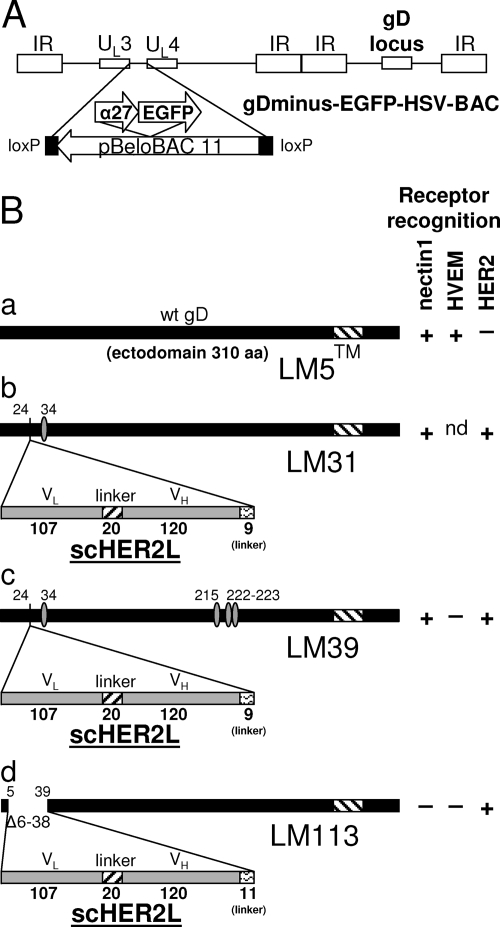
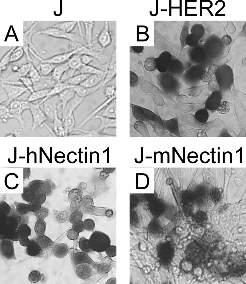
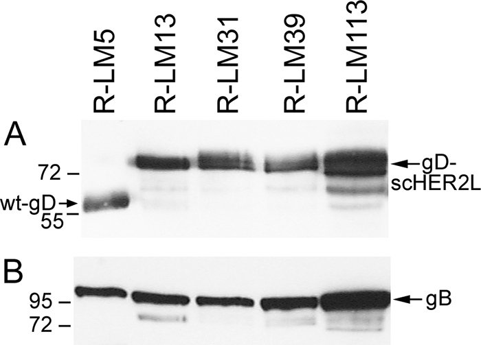
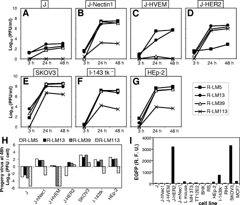
A novel frontier in the treatment of tumors that are difficult to treat is oncolytic virotherapy, in which a replication-competent virus selectively infects and destroys tumor cells. Herpes simplex virus (HSV) represents a particularly attractive system. Effective retargeting to tumor-specific receptors has been achieved by insertion in gD of heterologous ligands. Previously, our laboratory generated an HSV retargeted to human epidermal growth factor receptor 2 (HER2), a receptor overexpressed in about one-third of mammary tumors and in some ovarian tumors. HER2 overexpression correlates with increased metastaticity and poor prognosis. Because HER2 has no natural ligand, the inserted ligand was a single-chain antibody to HER2. The objective of this work was to genetically engineer an HSV that selectively targets the HER2-expressing tumor cells and that has lost the ability to enter cells through the natural gD receptors, HVEM and nectin1. Detargeting from nectin1 was attempted by two different strategies, point mutations and insertion of the single-chain antibody at a site in gD different from previously described sites of insertion. We report that point mutations at gD amino acids 34, 215, 222, and 223 failed to generate a nectin1-detargeted HSV. An HSV simultaneously detargeted from nectin1 and HVEM and retargeted to HER2 was successfully engineered by moving the site of single-chain antibody insertion at residue 39, i.e., in front of the nectin1-interacting surface and not lateral to it, and by deleting amino acid residues 6 to 38. The resulting recombinant, R-LM113, entered cells and spread from cell to cell solely via HER2.


Figures

FIG. 1.
FIG. 1.
(A) Schematic representation of the recombinant HSV-BACs generated in this study. The backbone of gD-minus-EGFP-HSV-BAC is shown as an example. The HSV-BACs are derived from pYEbac102 (54), which carries pBeloBAC11 sequences inserted between UL3 and UL4. In gD-minus-EGFP-HSV-BAC, the reporter cassette (α27-EGFP) is inserted in the BAC sequences. gD-minus-LacZ-HSV-BAC has the same structure but carries LacZ in place of EGFP. (B) Schematic representations of wt gD (a) and the gD-scHER2 chimeric proteins. gD of recombinant R-LM31 carries a substitution at amino acid residue 34 (b); gD of recombinant R-LM39 carries mutations at amino acid residues 34, 215, 222, and 223 (c); and gD of recombinant R-LM113 carries scHER2 in place of amino acid residues 6 to 38 (d). The boldface numbers indicate the length in amino acid residues of each fragment. The lightface numbers refer to amino acid residues according to wt gD coordinates. Mutated residues are indicated by ovals (Table 1). For each virus expressing wt or chimeric gD, the pattern of receptor recognition is summarized in the right-hand columns as +, −, or not determined (nd). TM, transmembrane domain. VH and VL, heavy- and light-chain variable domains of the anti-HER2 antibody 4D5; Δ, deletion. In panel B, the bars are drawn to scale.
FIG. 2.
FIG. 2.
The recombinant virus R-LM31 is not detargeted from the nectin1 receptor. Shown are micrographs of receptor-negative J cells (A) and J-HER2 (B), J-hNectin1 (C), and J-mNectin1 (D) expressing human HER2 and human or murine nectin1, respectively, exposed to R-LM31 at 10 PFU/cell. Infection was monitored as β-galactosidase activity by in situ X-Gal (5-bromo-4-chloro-3-indolyl-β-d-galactopyranoside) staining 16 h after infection.
FIG. 3.
FIG. 3.
Electrophoretic mobility of chimeric scHER2-gDs (A) or gB (B) expressed in SKOV3 cells infected with R-LM5, R-LM13, R-LM39, or R-LM113. The infected cell lysates were separated by sodium dodecyl sulfate-polyacrylamide gel electrophoresis, transferred to nitrocellulose membranes, and visualized by Western blotting with MAb BD80 against the C-terminal portion of the gD ectodomain (A) or MAb H1817 against gB (B), followed by peroxidase-conjugated anti-mouse IgG and enhanced chemiluminescence. In the recombinants, the insertion of scHER2L in gD resulted in slower electrophoretic mobility. The numbers on the left represent the migration positions of molecular mass markers (in kilodaltons).
FIG. 4.
FIG. 4.
Growth of R-LM39 and R-LM113 recombinants and of control viruses R-LM5 and R-LM13. (A to G) Replicate cultures of J (A), J-Nectin1 (B), J-HVEM (C), J-HER2 (D), SKOV3 (E), I-143 tk (F), and HEp-2 (G) cells were infected with R-LM5, R-LM13, R-LM39, or R-LM113 at 1 PFU/cell. Progeny virus was harvested at 3, 24, and 48 h after infection and titrated in SKOV3 cells. (H) Viral growth values at 48 h. (I) Infection of an array of cell lines with R-LM113. Monolayers were infected at 5 PFU/cell. Infection was quantified at 24 h as EGFP expression by means of a fluorometer. R.F.U., relative fluorescence units.
FIG. 5.
FIG. 5.
Blocking of infection of SKOV3 cells with R-LM39 (A) or R-LM113 (B) by antibodies to HER2 (Herceptin) or nectin1 (R1.302). SKOV3 cells were preincubated with the indicated concentrations of purified IgG from Herceptin, R1.302, the combination of Herceptin plus R1.302 or irrelevant mouse IgGs for 2 h at 4°C. For the combination of the two antibodies, the figures on the x axis indicate the concentration of each antibody. Virus was added to the antibody-containing medium and allowed to adsorb to the cells for 90 min at 4°C. Infection was monitored 16 h later as EGFP expression. One hundred percent represents the EGFP readings in untreated virus-infected cultures.
FIG. 6.
FIG. 6.
(A) Inhibition of cell-to-cell spread by Herceptin. SKOV3 cells infected with serial dilutions of the indicated viruses were overlaid with medium containing 1% SeaPlaque agarose ± 10 μg/ml Herceptin. Individual plaques were photographed at 48 h, and the plaque areas were measured by means of the Photoshop histogram tool and expressed as pixels. The histograms represent averages; the error bars represent standard deviations. (B) Representative plaques.

References

    1. Adler, H., M. Messerle, M. Wagner, and U. H. Koszinowski. 2000. Cloning and mutagenesis of the murine gammaherpesvirus 68 genome as an infectious bacterial artificial chromosome. J. Virol. 746964-6974. - PMC - PubMed
    1. Advani, S. J., R. R. Weichselbaum, R. J. Whitley, and B. Roizman. 2002. Friendly fire: redirecting herpes simplex virus-1 for therapeutic applications. Clin. Microbiol. Infect. 8551-563. - PubMed
    1. Aghi, M., and R. L. Martuza. 2005. Oncolytic viral therapies—the clinical experience. Oncogene 247802-7816. - PubMed
    1. Andreansky, S., L. Soroceanu, E. R. Flotte, J. Chou, J. M. Markert, G. Y. Gillespie, B. Roizman, and R. J. Whitley. 1997. Evaluation of genetically engineered herpes simplex viruses as oncolytic agents for human malignant brain tumors. Cancer Res. 571502-1509. - PubMed
    1. Andreansky, S. S., B. He, G. Y. Gillespie, L. Soroceanu, J. Markert, J. Chou, B. Roizman, and R. J. Whitley. 1996. The application of genetically engineered herpes simplex viruses to the treatment of experimental brain tumors. Proc. Natl. Acad. Sci. USA 9311313-11318. - PMC - PubMed

Publication types

MeSH terms