Review
. 2008 Aug 15;112(4):946-56.
doi: 10.1182/blood-2007-11-078097.

Mast cells and mastocytosis

Affiliations
Review

Mast cells and mastocytosis

Dean D Metcalfe. Blood. .

Abstract

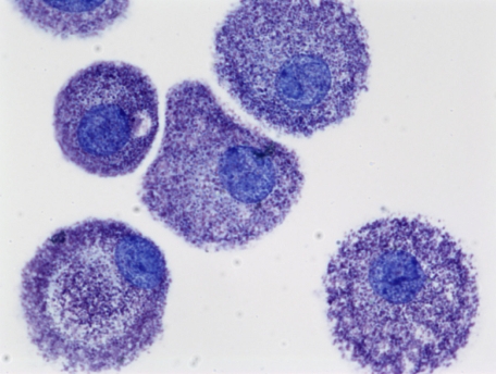
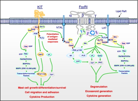
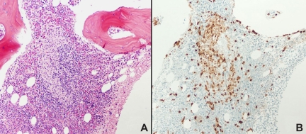
Mast cells have been recognized for well over 100 years. With time, human mast cells have been documented to originate from CD34+ cells, and have been implicated in host responses in both innate and acquired immunity. In clinical immunology, they are recognized for their central role in IgE-mediated degranulation and allergic inflammation by virtue of their expression of the high-affinity receptor for IgE and release of potent proinflammatory mediators. In hematology, the clinical disease of mastocytosis is characterized by a pathologic increase of mast cells in tissues, often associated with mutations in KIT, the receptor for stem cell factor. More recently, and with increased understanding of how human mast cells are activated through receptors including the high-affinity receptor for IgE and KIT, specific tyrosine kinase inhibitors have been identified with the potential to interrupt signaling pathways and thus limit the proliferation of mast cells as well as their activation through immunoglobulin receptors.


Figures

None
Dean D. Metcalfe
Figure 1
Figure 1
Human mast cells cultured from peripheral blood in SCF and stained with toluidine blue (cytoprep; 400×). The figure is provided courtesy of Madeleine K. Radinger (Laboratory of Allergic Diseases, National Institute of Allergy and Infectious Diseases, National Institutes of Health (LAD/NIAID/NIH). Images were obtained via digital microscopy using a Zeiss Axiophot (Jena, Germany) equipped with a Plan-Apochramat 100×/1.4 numeric aperature (NA) objective. Images were processed using Adobe Photoshop version 3.0 (Adobe Systems, San Jose, CA).
Figure 2
Figure 2
Signal transduction events initiated by Kit and FcϵRI leading to specific mast cell responses, and the integration of these pathways for the synergistic enhancement of mast cell mediator release. Dimerization of Kit, following ligation by SCF, results in activation of its intrinsic tyrosine kinase activity and autophosphorylation of specific tyrosine residues on its cytosolic tail. These phosphorylated residues provide docking sites for critical signaling molecules including Src kinases, Shc, phosphoinositide 3-kinase (PI3K), and phospholipase Cγ (PLCγ). Subsequent activation of the Ras-Raf-Map kinase (MAPK) cascade via the GTP exchanger Sos, PI3K, PLCγ, and JAK2 leads to the enhanced concentrations of Ca2+, and activation of transcription factors and survival pathways necessary for mast cell growth, differentiation, survival, cell migration, adhesion, and cytokine production. The Src kinases Lyn and Fyn may also contribute to some of these responses. Aggregation of FcϵRI, following binding of antigen to antigen-specific IgE molecules occupying the FcϵRI, results in recruitment of the tyrosine kinase Syk to the γ chain-ITAMs following translocation to the lipid raft microdomains and phosphorylation of specific tyrosines within these motifs by Lyn. This results in activation of Syk and the phosphorylation of the transmembrane adaptor molecules LAT and NTAL that coordinate downstream signaling by providing multiple phosphotyrosine-based docking sites for associating molecules such as PLCγ1 and the cytosolic adaptors Grb2, Gads, and Shc. These events regulate the activation of PLCγ1 and PLCγ2, which are required for calcium mobilization and PKC activation, essential signals for mast cell mediator release. A parallel pathway initiated by the tyrosine kinase, Fyn, leads to the activation of PI3K via the phosphorylation of the cytosolic adaptor Gab2. These events are also required for optimal degranulation and cytokine production, likely as a consequence of PI3K-dependent membrane association of the tyrosine kinase kinase, Btk, and activation of transcription factors. PI3K may also regulate the activation of sphingosine kinase (SK) and PLD, which produce the lipid mediators sphingosine-1-phosphate (S-1-P) and diacylglycerol (DAG), which also contribute to the critical calcium signal and PKC activation regulating mast cell mediator release. Concurrent with these events, the GTP exchangers Sos and Vav activate the Ras-Raf-MAPK pathway, which in turn contributes to the activation of specific transcription factors required for cytokine production. In addition, the MAPKs ERK1/2 control the activation of PLA2, which liberates arachidonic acid from membrane lipids for the subsequent generation of eicosanoids. NTAL appears to be a point of integration between the 2 pathways, providing an amplification mechanism also requiring PI3K and BTK, for the potentiation of antigen-dependent mediator release by SCF. Note that, although not depicted as such for clarity, many signaling events occur within specific microdomains within the cytosolic membrane. In addition, for clarity, several of the intermediary steps have been simplified and other pathways that may down-regulate mast cell activation have not been included. The figure and legend are provided courtesy of Alasdair M. Gilfillan (LAD/NIAID/NIH).
Figure 3
Figure 3
Bone marrow biopsy from a patient with systemic mastocytosis. Hematoxylin and eosin staining is shown in panel A, tryptase staining in panel B. Magnification ×100. Note the focal collections of mast cells, major criteria for the diagnosis of systemic mastocytosis. Courtesy of Irina Maric (NIH/Clinical Center, Department of Laboratory Medicine). Images were obtained via digital microscopy using an Olympus BH-2 microscope (Olympus, Melville, NY) equipped with a DPlan 10×/0.65 NA objective. Images were captured using an Olympus DP12 digital camera system and recorded on a 3.3V SmartMedia SSFDC card. Imaging software was Adobe Photoshop version 6.0 (Adobe Systems, San Jose, CA).

References

    1. Cavalcante M, Allodi S, Valente A, et al. Occurrence of heparin in the invertebrate styela plicata (tunicata) is restricted to cell layers facing the outside environment. J Biol Chem. 2000;275:36189–36196. - PubMed
    1. Malone DG, Metcalfe DD. Demonstration and characterization of a transient arthritis in rats following sensitization of synovial mast cells with antigen-specific IgE and parenteral challenge with specific antigen. Arthritis Rheum. 1998;31:1063–1067. - PubMed
    1. Nigrovic PA, Binstadt BA, Monach PA, et al. Mast cells contribute to initiation of autoantibody-mediated arthritis via lL-1. Proc Natl Acad Sci U S A. 2007;104:2325–30. - PMC - PubMed
    1. Brown JM, Swindle EJ, Kushnir-Sukhov NM, Holian A, Metcalfe DD. Silica directed mast cell activation is enhanced by scavenger receptors. Am J Respir Cell Mol Biol. 2007;36:43–52. - PMC - PubMed
    1. Ehrlich P. Leipzig, Germany: University of Leipzig; 1878. Beitrage zur theorie und praxis der histologischen färbung [thesis].

Publication types