. 2008 Aug 15;181(4):2277-84.
doi: 10.4049/jimmunol.181.4.2277.

Retinoic acid increases Foxp3+ regulatory T cells and inhibits development of Th17 cells by enhancing TGF-beta-driven Smad3 signaling and inhibiting IL-6 and IL-23 receptor expression

Affiliations

Retinoic acid increases Foxp3+ regulatory T cells and inhibits development of Th17 cells by enhancing TGF-beta-driven Smad3 signaling and inhibiting IL-6 and IL-23 receptor expression

Sheng Xiao et al. J Immunol. .

Abstract

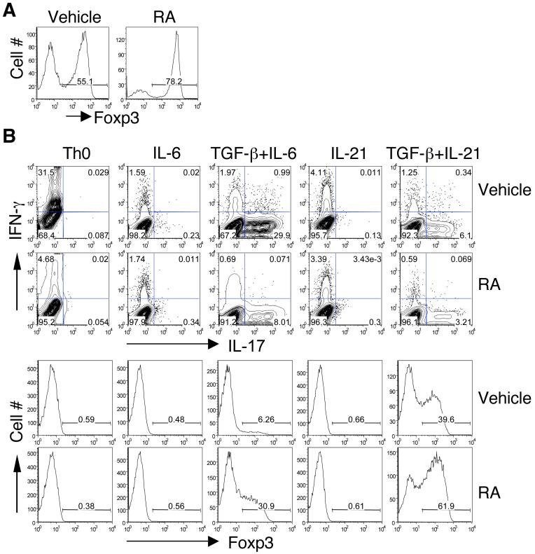
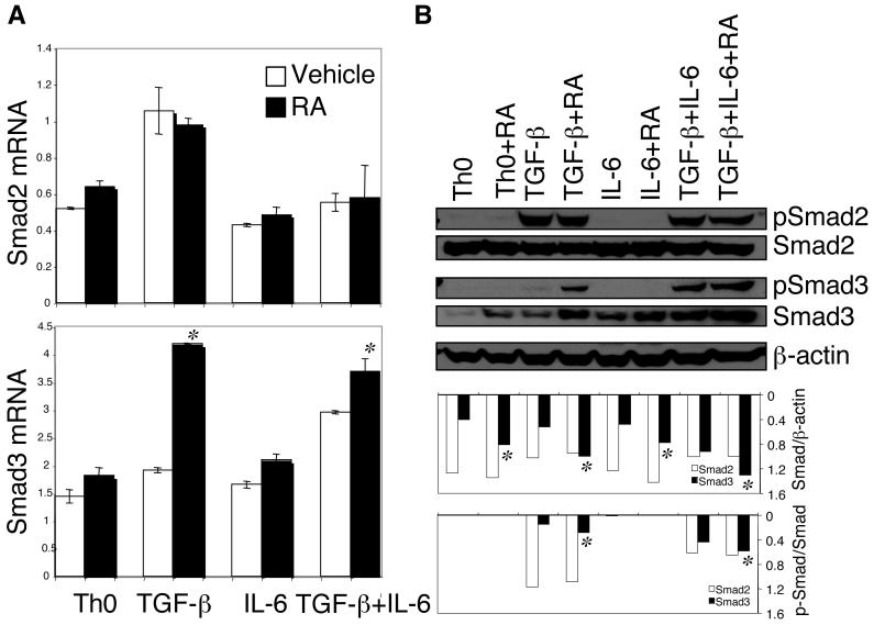
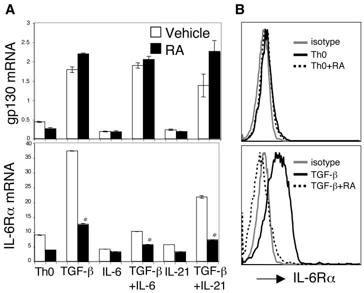
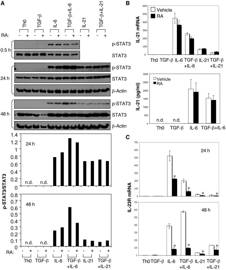
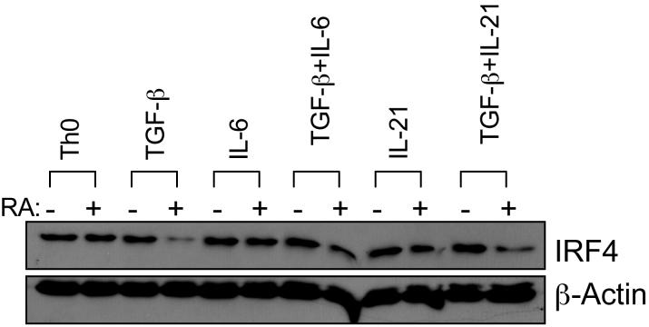
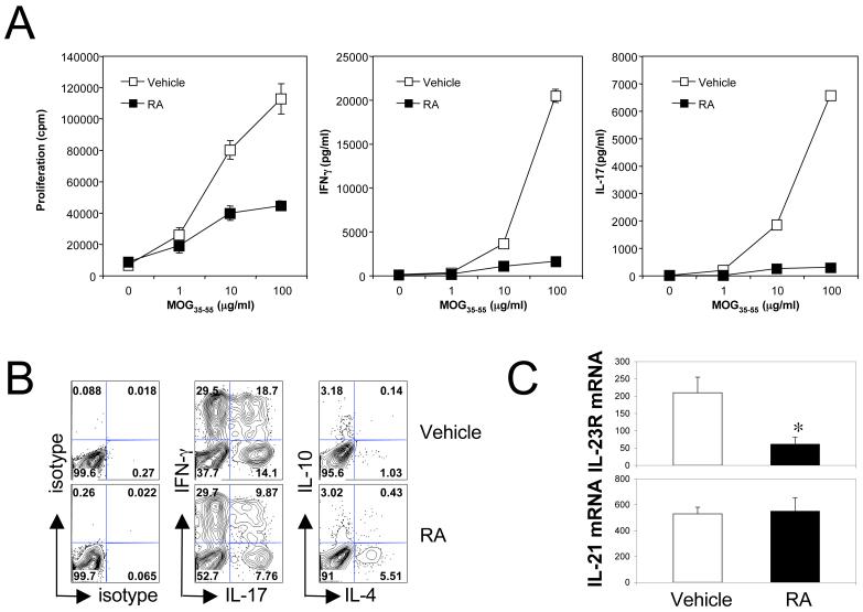
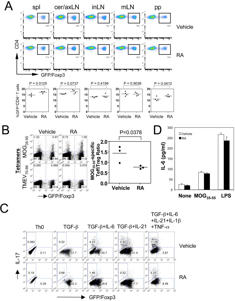
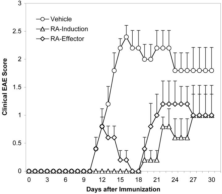
The de novo generation of Foxp3+ regulatory T (Treg) cells in the peripheral immune compartment and the differentiation of Th17 cells both require TGF-beta, and IL-6 and IL-21 are switch factors that drive the development of Th17 cells at the expense of Treg cell generation. The major vitamin A metabolite all-trans retinoic acid (RA) not only enforces the generation of Treg cells but also inhibits the differentiation of Th17 cells. Herein we show that RA enhances TGF-beta signaling by increasing the expression and phosphorylation of Smad3, and this results in increased Foxp3 expression even in the presence of IL-6 or IL-21. RA also inhibits the expression of IL-6Ralpha, IRF-4, and IL-23R and thus inhibits Th17 development. In vitro, RA significantly promotes Treg cell conversion, but in vivo during the development of experimental autoimmune encephalomyelitis it does not increase the frequency of Treg cells in the face of an ongoing inflammation. However, RA suppresses the disease very efficiently by inhibiting proinflammatory T cell responses, especially pathogenic Th17 responses. These data not only identify the signaling mechanisms by which RA can affect both Treg cell and Th17 differentiation, but they also highlight that in vivo during an autoimmune reaction, RA suppresses autoimmunity mainly by inhibiting the generation of effector Th17 cells.


Figures

Figure 1
Figure 1. Differential effect of RA in the development of Foxp3+ Treg and Th17 cells
Naïve CD4+ T cells were activated with anti-CD3 and anti-CD28 for 4 days under indicated conditions in the absence or presence of RA or Vehicle (DMSO). Aliquots of cells cultured with TGF-β (A) or different combinations of cytokines (B) were fixed, permeabilized, and stained intracellularly with PEFoxp3 mAb and analyzed by flow cytometry. Cells were also reactivated with PMA and ionomycin for 4 h in the presence of GolgiStop, and then stained with APC-IFN-g and PE-IL-17 mAb and analyzed by flow cytometry. Numbers in lower right quadrants indicate the percentage of IL-17-producing cells (B). Data are representative of 3-5 experiments.
Figure 2
Figure 2. RA enhanced TGF-β-induced expression and phosphorylation of Smad3 but not Smad2
Naïve CD4+ T cells were activated with anti-CD3 and anti-CD28 for 24 h under indicated conditions with or without RA. (A) Total RNA was isolated and expression of Smad2 and Smad3 mRNA was determined by Taqman quantitative PCR. (B) Cell lysates from indicated conditions were separated by SDS-PAGE gel, and total Smad and p-Smad were detected by immunoblotting. For quantification, the ratio of total Smad/β-actin and the ratio of p-Smad/total Smad were determined. Data are representative of three experiments. *, P <; 0.05 when RA treatment was compared to vehicle treatment.
Figure 3
Figure 3. RA blocked TGF-β-induced upregulation of IL-6Rα expression
Naïve CD4+ T cells were activated under indicated conditions with or without RA. After 48 h, cells were collected for the determination of gp130 and IL-6Rα mRNA by Taqman quantitative PCR (A) and surface expression of IL-6Ra protein by flow cytometry (B). Data are representative of three experiments. *, P < 0.05 when RA treatment compared to vehicle treatment.
Figure 4
Figure 4. Effect of RA on STAT3 phosphorylation, IL-21 production, and IL-23R expression in CD4+ cells cultured with different combinations of TGF-β, IL-6, and IL-21.
Naïve CD4+ T cells were activated under indicated conditions with or without RA. Cells were collected for the determination of STAT3 phosphorylation by immunoblotting at various time points, and the ratio of p-STAT3/total STAT3 was analyzed (A). IL-21 mRNA expression at 24 h by Taqman quantitative PCR and IL-21 production in 96-h culture supernatants by ELISA (B). Expression of IL-23R mRNA at 24 h and 48 h by Taqman quantitative PCR (C). Data are representative of 3-5 experiments. *, P < 0.05 when RA treatment was compared to vehicle treatment.
Figure 5
Figure 5. RA decreased expression of IRF4 protein in the presence of TGF-β, IL-6 or IL-21
Naïve CD4+ T cells were activated under indicated conditions with or without RA. Cells were collected after 24 h or 48 h, lyzed, and detected for IRF4 protein expression by immunoblotting. Data are representative of three experiments.
Figure 6
Figure 6. Treatment with RA decreased pathogenic Th17 and Th1 responses and IL-23R expression in CD4+ T cells
C57BL/6 mice were immunized with MOG35-55/CFA, and RA was intraperitoneally injected every other day from day 0 to day 8. On day 10, (A) draining lymph node cells were isolated and restimulated with MOG35-55. Proliferation and cytokine production in 48-h culture supernatants were measured by 3[H]-thymidine incorporation and ELISA assays, respectively; (B) isolated spleen cells were cultured with 5 μg/ml of MOG35-55 for 5 days. Cytokine production from CD4+ cells was determined by intracellular cytokine staining following 4-h activation with PMA and ionomycin; (C) expression of IL-23R and IL-21 mRNA was measured in CD4+ T cells purified from spleen cells by Taqman quantitative PCR. *, P < 0.05 when RA treatment was compared to vehicle treatment. Data are the average or representative of 4-6 mice in each group.
Figure 7
Figure 7. RA in vivo did not measurably increase the population of Foxp3+ Treg cells
Foxp3gfp.KI mice were immunized with MOG35-55/CFA, and RA was intraperitoneally injected every other day from day 0 to day 8. On day 10, (A) the frequency of GFP+ Treg cells was determined in different lymphoid compartments by flow cytometry. “Spl”, spleen; “cer/ax”, cervical/axillary; “in”, inguinal; “m”, mesenteric; “pp”, peyer's patch; (B) spleen cells were restimulated with 2 μg/ml of MOG35-55 for 5 days, and then stained with PE-conjugated IAb tetramer, APC-anti-CD4, and 7-AAD. The MOG35-55/IAb tetramer-positive cells were determined in the live CD4+ cell population (CD4+7-AAD-). TMEV70-86/IAS tetramers were used as a negative control. (C) Inflammatory cytokines IL-6, TNF-α, and IL-1β inhibited RA-enhanced Foxp3 expression but could not rescue RA-inhibited IL-17 production. Naïve CD4+ T cells (CD4+ CD62Lhi Foxp3/GFP-) purified from Foxp3gfp.KI mice were activated with anti-CD3 and anti-CD28 under indicated conditions in the absence or presence of RA for 5 days. Cells were reactivated with PMA and ionomycin for 4 h in the presence of GolgiStop. Then, cells were stained with APC-anti-CD4, PE-anti-IL-17 and 7-AAD, and analyzed by flow cytometry for IL-17 production and Foxp3/GFP expression in gated CD4+7-AAD- cells. (D) Isolated spleen cells from immunized mice on day 10 were cultured with 5 μmg/ml of MOG35-55 peptide or 500ng/ml LPS. IL-6 production was determined in 48-h cultures by ELISA. None, no treatment.
Figure 8
Figure 8. Treatment with RA inhibited EAE both during the induction and after disease onset
C57BL/6 mice were immunized with MOG35-55/CFA, and RA was intraperitoneally injected every other day starting from day 0 (RA-induction) or after all mice in the group showed signs of clinical EAE (RA-effector). Mice were evaluated daily for the signs of EAE. EAE was significantly (P < 0.05) reduced in mice in the “RA-induction” group after day 10 and in the “RA-effector” group after day12 compared to the vehicle treated mice.

References

    1. Abbas AK, Murphy KM, Sher A. Functional diversity of helper T lymphocytes. Nature. 1996;383:787. - PubMed
    1. Harrington LE, Mangan PR, Weaver CT. Expanding the effector CD4 T-cell repertoire: the Th17 lineage. Curr Opin Immunol. 2006;18:349. - PubMed
    1. Bettelli E, Oukka M, Kuchroo VK. T(H)-17 cells in the circle of immunity and autoimmunity. Nat Immunol. 2007;8:345. - PubMed
    1. Korn T, Oukka M. Dynamics of antigen-specific regulatory T-cells in the context of autoimmunity. Semin Immunol. 2007;19:272. - PubMed
    1. Oukka M. Interplay between pathogenic Th17 and regulatory T cells. Ann Rheum Dis. 2007;66(Suppl 3):iii87. - PMC - PubMed

Publication types

MeSH terms