. 2008 Oct;295(4):C966-74.
doi: 10.1152/ajpcell.00008.2008. Epub 2008 Aug 6.

Connexin26 deafness associated mutations show altered permeability to large cationic molecules

Affiliations

Connexin26 deafness associated mutations show altered permeability to large cationic molecules

Gülistan Meşe et al. Am J Physiol Cell Physiol. 2008 Oct.

Abstract

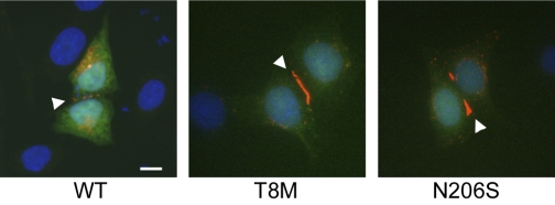
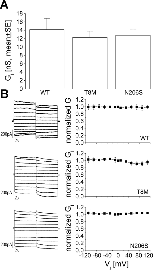
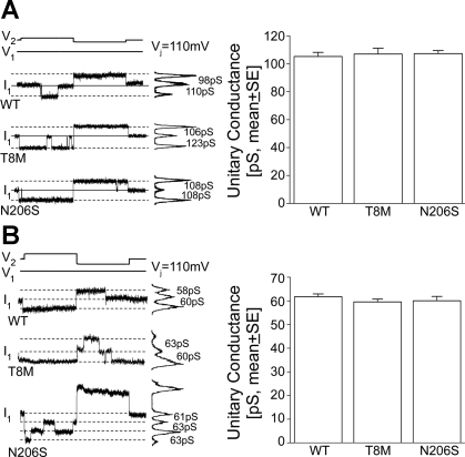
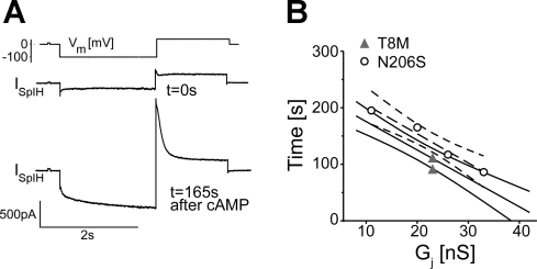
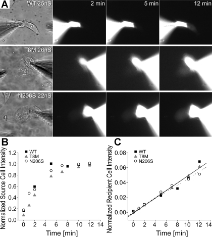
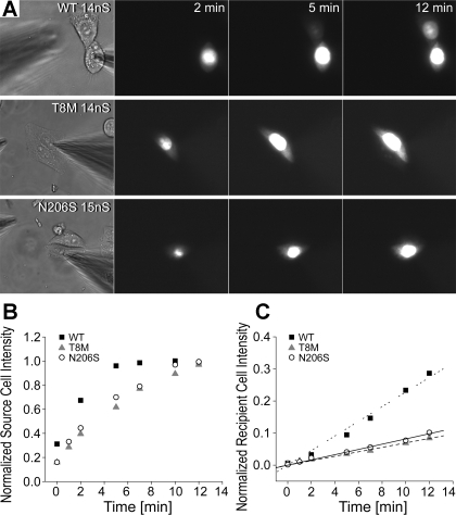
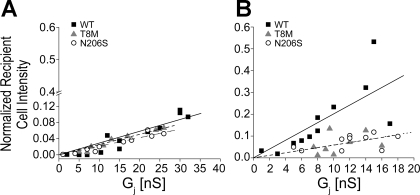
Intercellular communication is important for cochlear homeostasis because connexin26 (Cx26) mutations are the leading cause of hereditary deafness. Gap junctions formed by different connexins have unique selectivity to large molecules, so compensating for the loss of one isoform can be challenging in the case of disease causing mutations. We compared the properties of Cx26 mutants T8M and N206S with wild-type channels in transfected cells using dual whole cell voltage clamp and dye flux experiments. Wild-type and mutant channels demonstrated comparable ionic coupling, and their average unitary conductance was approximately 106 and approximately 60 pS in 120 mM K(+)-aspartate(-) and TEA(+)-aspartate(-) solution, respectively, documenting their equivalent permeability to K(+) and TEA(+). Comparison of cAMP, Lucifer Yellow (LY), and ethidium bromide (EtBr) transfer revealed differences in selectivity for larger anionic and cationic tracers. cAMP and LY permeability to wild-type and mutant channels was similar, whereas the transfer of EtBr through mutant channels was greatly reduced compared with wild-type junctions. Altered permeability of Cx26 to large cationic molecules suggests an essential role for biochemical coupling in cochlear homeostasis.


Figures

Fig. 1.
Fig. 1.
Connexin 26 (Cx26) wild-type and NH2-terminal mutant Thr8Met (T8M) and transmembrane domain mutant Asn206Ser (N206S) protein expression and localization are shown. HeLa cells were transiently transfected with wild-type, T8M, and N206S constructs and were examined by immunostaining and fluorescence microscopy. Cells individually expressed wild-type and mutant T8M and N206S proteins and properly targeted them to the cell membrane especially at the region of cell-to-cell apposition. Arrowheads point to the punctate staining of Cx26 gap junction plaques at cell-cell contact areas. Green represents green fluorescent protein (GFP), blue shows DAPI staining of cell nuclei, and red is Cy3 staining of Cx26 protein. Scale bar = 10 μm.
Fig. 2.
Fig. 2.
Conductance and voltage-gating properties of wild-type, T8M, and N206S channels. A: comparison of macroscopic junctional conductance of Cx26 wild-type (n = 15), T8M (n = 16), and N206S channels (n = 20) in transiently transfected HeLa cells showed that they were not statistically different from each other (ANOVA, P < 0.05). Data are means ± SE. B: junctional currents from HeLa cell pairs transfected with wild-type Cx26, T8M, and N206S were recorded during application of a series of transjunctional voltages (Vjs) ranging from +110 to −110 mV in 20-mV increments (left). The normalized junctional conductance (means ± SE) versus Vjs was plotted (right). Cx26 wild-type and mutant T8M and N206S channels demonstrated very little voltage dependence.
Fig. 3.
Fig. 3.
Single channel properties of wild-type, T8M, and N206S channels. Single channel currents recorded from cell pairs expressing wild-type, T8M, and N206S at a Vj of ±110 mV in transiently transfected Neuro-2A cells. A: current histograms of the single channels for wild-type and mutant connexins revealed unitary conductances between 98 and 123 pS in 120 mM K+-aspartate (left). Solid line represents the zero junctional current and the dashed lines indicate open-state currents. The averaged unitary conductances of wild-type, T8M, and N206S channels were statistically indistinguishable from each other (ANOVA, P < 0.05) (right). B: with the use of 120 mM TEA+-aspartate pipette solution (left), Cx26 wild-type, T8M, and N206S channels had unitary conductances between 58 and 63 pS and the mean unitary conductances for all three channels were comparable to each other (ANOVA, P < 0.05, right). Data represented as means ± SE.
Fig. 4.
Fig. 4.
Comparison of cAMP permeability through Cx26 wild-type, T8M, and N206S. A: SpIH current trace at the beginning of cAMP injection into source cell at t = 0 s and at t = 165 s when the current was saturated due to cAMP transfer into recipient cell from the injected cell, showing the SpIH-dependent current increase. B: SpIH saturation time for each channel was plotted against junctional conductance. Dark straight line represents our previously published SpIH activation times for wild-type channels (23), and light solid lines are the 95% confidence interval. Dashed line is the first-order regression for N206S channels, and the curved dashed lines are the 95% confidence interval. The SpIH activation times for N206S channels were not significantly different from wild-type and T8M channels whose saturation times overlap with wild-type Cx26.
Fig. 5.
Fig. 5.
Permeability of wild-type and mutant channels to Lucifer yellow (LY). Measurements of LY flux from cell pairs expressing wild-type Cx26, T8M, and N206S followed by measurements of junctional conductance (gj). A: epifluorescent micrographs taken at 2, 5, and 12 min for cell pairs of similar junctional conductance (22–26 nS) demonstrated progressive fluorescent intensity increases in the recipient cells for each channel type. B: fluorescent intensities of the source cells of wild-type, T8M, and N206S were normalized to the maximum fluorescent value in the source cells and was plotted as a function of time. For all three examples shown in A, the source cells had similar loading patterns. C: normalized intensities of recipient cells expressing wild-type, T8M, and N206S showed a time-dependent increase in the intensities of recipient cells over the course of 12 min, implicating dye transfer from the source cells to the recipient cells.
Fig. 6.
Fig. 6.
Permeability of wild-type and mutant channels to ethidium bromide (EtBr). EtBr transfer through cells expressing the wild-type and mutant channels was compared between pairs with similar junctional conductance. A: examples of epifluorescent micrographs (taken at 2, 5, and 12 min) of cell pairs expressing wild-type, T8M and N206S proteins during EtBr flux experiments. Transfer of EtBr through wild-type Cx26 channels was readily observed after the opening of the patch seal in the source cell, as seen by the increased intensity of the recipient cell. There was little visible fluorescent intensity in the recipient cells coupled by T8M and N206S channels. B: normalized fluorescent intensities of source cells expressing wild-type, T8M, and N206S Cx26 in A were plotted as a function of time (minutes) to show dye injection into the cells. C: fluorescent intensity of the recipient cells was increased over time. Both the T8M and N206S mutant channels had greatly reduced permeability relative to wild-type channels.
Fig. 7.
Fig. 7.
Relative intensity of recipient cells for LY and EtBr as a function of junctional conductance (gj). The relative intensities of recipient cells of Cx26 wild-type, T8M, and N206S junctions in different cell pairs were analyzed at the 12-min time point and plotted against the gj. A: relationship between the relative intensity and the gj for wild-type, T8M, and N206S for LY was linear [R2 values were 0.90 (straight line), 0.96 (dashed line) and 0.92 (dotted line), respectively]. LY passage between pairs of cells expressing wild-type, T8M, and N206S was comparable to each other, and statistically were not different (ANOVA, P < 0.05). B: EtBr transfer between cell pairs expressing wild-type Cx26 showed a modest linear correlation between relative intensity of recipient cell and gj with a R2 value of 0.92 (straight line). T8M and N206S channels showed only a weak correlation between gj versus relative intensity of recipient cell and greatly reduced EtBr permeability. The slopes of fitted lines for T8M (dashed fitting line) and N206S (dotted fitting line) channels were 27.6% and 28% of that of wild-type Cx26.

References

    1. Ahmad S, Tang W, Chang Q, Qu Y, Hibshman J, Li Y, Sohl G, Willecke K, Chen P, Lin X. Restoration of connexin26 protein level in the cochlea completely rescues hearing in a mouse model of human connexin30-linked deafness. Proc Natl Acad Sci USA 104: 1337–1341, 2007. - PMC - PubMed
    1. Barrio LC, Suchyna T, Bargiello T, Xu LX, Roginski RS, Bennett MV, Nicholson BJ. Gap junctions formed by connexins 26 and 32 alone and in combination are differently affected by applied voltage. Proc Natl Acad Sci USA 88: 8410–8414, 1991. - PMC - PubMed
    1. Beahm DL, Oshima A, Gaietta GM, Hand GM, Smock AE, Zucker SN, Toloue MM, Chandrasekhar A, Nicholson BJ, Sosinsky GE. Mutation of a conserved threonine in the third transmembrane helix of alpha- and beta-connexins creates a dominant-negative closed gap junction channel. J Biol Chem 281: 7994–8009, 2006. - PubMed
    1. Beblo DA, Veenstra RD. Monovalent cation permeation through the connexin40 gap junction channel. Cs, Rb, K, Na, Li, TEA, TMA, TBA, and effects of anions Br, Cl, F, acetate, aspartate, glutamate, and NO3. J Gen Physiol 109: 509–522, 1997. - PMC - PubMed
    1. Beltramello M, Bicego M, Piazza V, Ciubotaru CD, Mammano F, D'Andrea P. Permeability and gating properties of human connexins 26 and 30 expressed in HeLa cells. Biochem Biophys Res Commun 305: 1024–1033, 2003. - PubMed

Publication types