. 2008 Sep;36(16):5281-9.
doi: 10.1093/nar/gkn507. Epub 2008 Aug 6.

Tetrahymena thermophila and Candida albicans group I intron-derived ribozymes can catalyze the trans-excision-splicing reaction

Affiliations

Tetrahymena thermophila and Candida albicans group I intron-derived ribozymes can catalyze the trans-excision-splicing reaction

P Patrick Dotson 2nd et al. Nucleic Acids Res. 2008 Sep.

Abstract

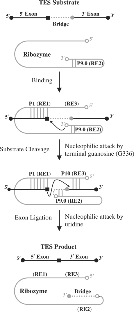
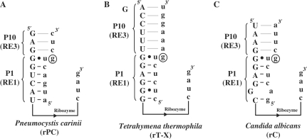
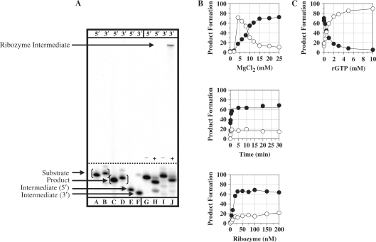
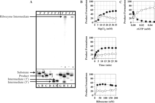
Group I intron-derived ribozymes can catalyze a variety of non-native reactions. For the trans-excision-splicing (TES) reaction, an intron-derived ribozyme from the opportunistic pathogen Pneumocystis carinii catalyzes the excision of a predefined region from within an RNA substrate with subsequent ligation of the flanking regions. To establish TES as a general ribozyme-mediated reaction, intron-derived ribozymes from Tetrahymena thermophila and Candida albicans, which are similar to but not the same as that from Pneumocystis, were investigated for their propensity to catalyze the TES reaction. We now report that the Tetrahymena and Candida ribozymes can catalyze the excision of a single nucleotide from within their ribozyme-specific substrates. Under the conditions studied, the Tetrahymena and Candida ribozymes, however, catalyze the TES reaction with lower yields and rates [Tetrahymena (k(obs)) = 0.14/min and Candida (k(obs)) = 0.34/min] than the Pneumocystis ribozyme (k(obs) = 3.2/min). The lower yields are likely partially due to the fact that the Tetrahymena and Candida catalyze additional reactions, separate from TES. The differences in rates are likely partially due to the individual ribozymes ability to effectively bind their 3' terminal guanosines as intramolecular nucleophiles. Nevertheless, our results demonstrate that group I intron-derived ribozymes are inherently able to catalyze the TES reaction.


Figures

Figure 1.
Figure 1.
Diagram of the ribozyme-mediated trans-excision-splicing reaction. The P. carinii ribozyme is denoted in gray and the 5′- and 3′-exon sequences are denoted by black lines. The black square within the 5′-exon region represents a uridine and the gray circle adjacent to the 3′-exon region represents a guanosine. The bridge region to be excised is denoted as a dotted line. The P1 helix is formed through base-pairing of recognition element 1 (RE1) with the 5′-exon. The P10 helix is formed through base-pairing of recognition element 3 (RE3) with the 3′-exon. The first step (substrate cleavage) is catalyzed by G336 (open circle at 3′-end of ribozyme) resulting in the covalent attachment of the 3′-end of the substrate to the 3′-end of the ribozyme. The second step (exon ligation) proceeds through attack of the uridine upon the guanosine of the substrate, resulting in ligation of the flanking sequences.
Figure 2.
Figure 2.
Diagram of P. carinii (rPC), T. thermophila (rT-X) and C. albicans (rC) ribozyme constructs. The sequence of the Pneumocystis (A), Tetrahymena (B) and Candida (C) ribozyme constructs are shown in uppercase letters with their respective substrate shown in lowercase letters. Only the sequences that are involved with binding the individual substrates are shown, as well as the 3′-end of the ribozymes. Note that the remaining ribozyme sequences, represented with a line, are different for each of the constructs. The nucleotide to be excised (a guanosine) is circled for all three ribozyme constructs.
Figure 3.
Figure 3.
TES reactions conducted with the P. carinii (rPC) ribozyme. (A) Polyacrylamide gel of TES reactions using either 5′ or 3′-end radiolabeled substrates. Reactions were conducted using 100 nM rPC, H0Mg (−) or H15Mg (+) buffer for 30 min at 44°C. Lanes A, C and E contain 5′-end labeled size controls, respectively. Lanes B, D and F contain 3′-end radiolabeled size controls, respectively. Note that the 3′-end labeled size controls are one nucleotide larger than 5′-end labeled size controls. Lanes G–J contain the rPC ribozyme with 5′-end radiolabeled (lanes G and H) or 3′-end radiolabeled (lanes I and J) substrate. (B) Graphs of TES reactions using 5′-end radiolabeled substrate. All reactions were conducted as above except for the changing variable. (C) Graph of exogenous guanosine concentration dependence for TES reactions. All data points represent the average of at least two independent reactions with standard deviations typically below 10%. For all graphs, the TES product is represented by filled circles and the substrate cleavage product is represented as open circles.
Figure 4.
Figure 4.
TES reactions conducted with the T. thermophila (rT-X) ribozyme. (A) Polyacrylamide gel of TES reactions using either 5′ or 3′-end radiolabeled substrates. Reactions were conducted using 166 nM rT-X, H0Mg (−) or H10Mg (+) buffer, for 45 min at 44°C. Lanes A, C and F contain 5′-end radiolabeled size controls, respectively. Lanes B, D and E contain 3′-end radiolabeled size controls, respectively. Note that the 3′-end radiolabeled size controls are one nucleotide larger than 5′-end radiolabeled size controls. Lanes G–J contain the normal rT-X ribozyme with 5′-end radiolabeled (lanes G and H) or 3′-end radiolabeled (lanes I and J) substrate. (B) Graphs of TES reactions using 5′-end radiolabeled substrate. All reactions were conducted as above except for the changing variable. (C) Graph of rGTP concentration dependence for TES reactions conducted with the rT-X ribozyme. All data points represent the average of at least two independent reactions with standard deviations typically below 10%. Note for all graphs, the TES product is represented by filled triangles and the substrate cleavage product is represented as open triangles.
Figure 5.
Figure 5.
TES reactions conducted with the C. albicans (rC) ribozyme. (A) Polyacrylamide gel of TES reactions using both 5′ and 3′-end radiolabeled substrates. Reactions were conducted using 75 nM rC, H0Mg (−) or H25Mg (+) buffer, for 30 min at 44°C. Lanes A, C and E contain 5′-end radiolabeled size controls, respectively. Lanes B, D and F contain 3′-end radiolabeled size controls, respectively. Note that the 3′-end radiolabeled size controls are one nucleotide larger than 5′-end radiolabeled size controls. Lanes G–J contain the normal rC ribozyme with 5′-end radiolabeled (lanes G and H) or 3′-end radiolabeled (lanes I and J) substrate. (B) Graphs of TES reactions using 5′-end radiolabeled substrate. All reactions were conducted as above except for the changing variable. (C) Graph of rGTP concentration dependence for TES reactions conducted with the rC ribozyme. All data points represent the average of at least two independent reactions, with standard deviations typically below 10%. Note for all graphs, the TES product is represented by filled triangles and the substrate cleavage product is represented as open triangles.

References

    1. Cech TR. Self-splicing of group I introns. Annu. Rev. Biochem. 1990;59:543–568. - PubMed
    1. Dotson PP, II, Testa SM. Group I intron-derived ribozyme recombination reactions. Rec. Develop. Nucleic Acids Res. 2006;2:307–324.
    1. Bell MA, Johnson AJ, Testa SM. Ribozyme-catalyzed excision of targeted sequences from within RNAs. Biochemistry. 2002;41:15327–15333. - PubMed
    1. Dotson PP, II, Sinha J, Testa SM. A Pneumocystis carinii group I intron-derived ribozyme utilizes an endogenous guanosine as the first reaction step nucleophile in the trans excision-splicing reaction. Biochemistry. 2008;47:4780–4787. - PubMed
    1. Zaug AJ, Grosshans CA, Cech TR. Sequence-specific endoribonuclease activity of the Tetrahymena ribozyme: enhanced cleavage of certain oligonucleotide substrates that form mismatched ribozyme-substrate complexes. Biochemistry. 1988;27:8924–8931. - PubMed

Publication types