. 2008 Aug 6;28(32):8138-43.
doi: 10.1523/JNEUROSCI.1006-08.2008.

Developmental neuronal death in hippocampus requires the microglial CD11b integrin and DAP12 immunoreceptor

Affiliations

Developmental neuronal death in hippocampus requires the microglial CD11b integrin and DAP12 immunoreceptor

Shirley Wakselman et al. J Neurosci. .

Abstract

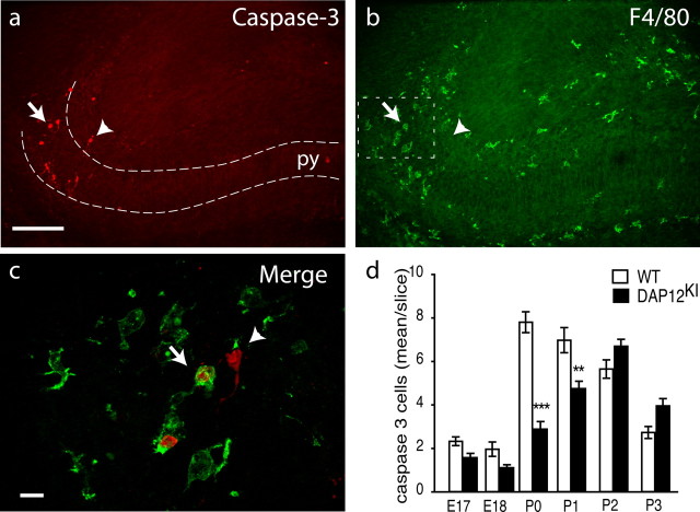
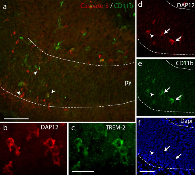
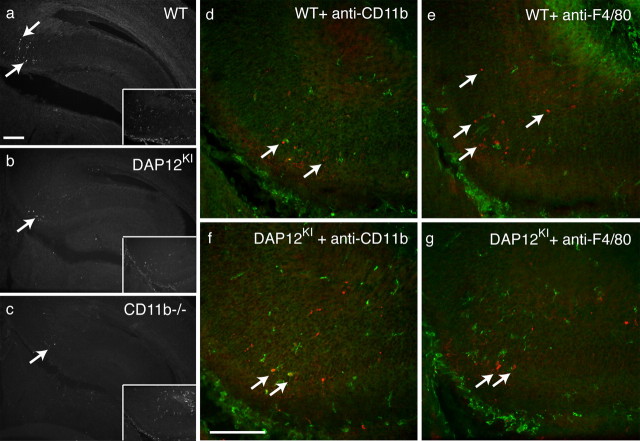
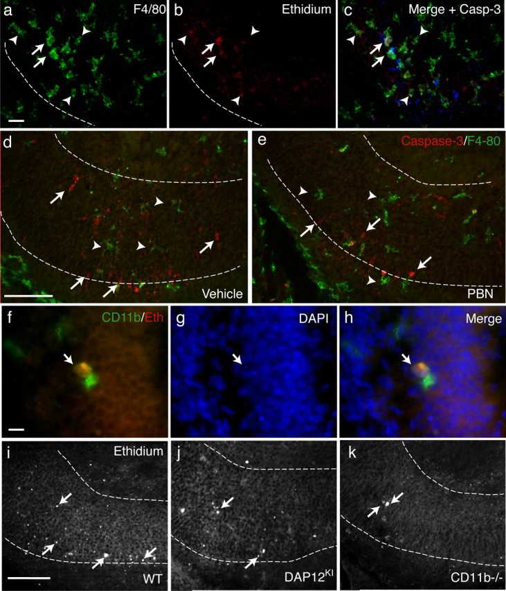
In several brain regions, microglia actively promote neuronal apoptosis during development. However, molecular actors leading microglia to trigger death remain mostly unknown. Here, we show that, in the developing hippocampus, apoptotic neurons are contacted by microglia expressing both the integrin CD11b and the immunoreceptor DAP12. We demonstrate that developmental apoptosis decreases in mice deficient for CD11b or DAP12. In addition, function-blocking antibodies directed against CD11b decrease neuronal death when injected into wild-type neonates, but have no effect when injected into DAP12-deficient littermates. This demonstrates that DAP12 and CD11b act in converging pathways to induce neuronal death. Finally, we show that DAP12 and CD11b control the production of microglial superoxide ions, which kill the neurons. Thus, our data show that the process of developmental neuronal death triggered by microglia is similar to the elimination of pathogenic cells by the innate immune cells.


Figures

Figure 1.
Figure 1.
Apoptotic neurons in neonate hippocampus are in contact with microglia. Double labeling of P0 hippocampus with antibodies against activated caspase-3 (red) (a) and F4/80 (green) (b). The arrow and arrowhead indicate caspase-3-positive cells that are apposed or not with microglia, respectively. Scale bar, 100 μm. py, Pyramidal cell layer. c, Higher magnification of the subiculum. Activated caspase-3-positive cells are surrounded (arrow) or contacted (arrowhead) by microglial processes. Scale bar, 10 μm. d, Quantification of developmental death in the hippocampus between E17 and P3 in WT (white bar) or in DAP12KI (black bar). Shown are mean ± SEM. ***p < 0.0001; **p < 0.001.
Figure 2.
Figure 2.
Microglia contacting apoptotic cells in the developing hippocampus express CD11b and DAP12 and TREM-2. a, Activated caspase-3-positive cells (red; arrowheads) are apposed to CD11b-positive microglia (green). py, Pyramidal cell layer. Scale bar, 50 μm. b, c, Double labeling of neonate hippocampus with anti-DAP12 antibody (b) and TREM-2 antibody (c). Scale bar, 20 μm. d–f, Triple labeling of P0 hippocampus with anti-DAP12 antibody (d), anti-CD11b antibody (e), and DAPI (f). The arrows indicate double-labeled microglia in contact with condensed nucleus. The arrowhead shows a condensed nucleus not in contact with labeled microglia. Scale bar, 50 μm.
Figure 3.
Figure 3.
Decreased developmental apoptosis on DAP12 or CD11b inactivation. Anti-activated caspase-3 immunoreactivity in the hippocampus of neonates: WT (a); DAP12KI (b); CD11b−/− (c). The inset shows the codetection of F4/80 immunoreactivity in the subicular region. d–g, Codetection of activated caspase-3-positive cells (red; arrows) and microglia (green) in the subicular region of WT (d, e) or DAP12KI (f, g) P1 mice injected 24 h earlier with rat anti-CD11b function-blocking antibody (d, f) or rat anti-F4/80b antibody (e, g). The green staining shows the detection of rat antibodies present in the tissue by labeling the slices with an anti-rat antibody coupled to Alexa 488 fluorophore without previous incubation with primary antibodies. Scale bars, 100 μm.
Figure 4.
Figure 4.
DAP12- and CD11b-dependent production of superoxide ions induces the developmental death of neurons. a–c, In the subiculum of WT neonate, triple detection of microglia by F4/80 immunoreactivity (a); cells producing superoxide ions revealed by the oxidation of DHE into red-fluorescent ethidium (b). c, Merge image with activated caspase-3 staining (blue). Some ethidium-producing microglia contact apoptotic cells (arrows) but most do not (arrowheads). Scale bar, 20 μm. d, e, Double detection of caspase-3 labeling (red) and F4/80 immunoreactivity (green) in hippocampus on systemic injection of vehicle (d) or free radical scavenger PBN (e). Scale bar, 50 μm. f, High magnification of a CD11b-positive microglia (green) that has accumulated ethidium (red). Scale bar, 10 μm. g, DAPI staining revealed condensed nucleus (arrow) of apoptotic cells. h, Merge image showing a contact between superoxide producing CD11b-positive microglia and the apoptotic nucleus. i–k, Oxidation of DHE in WT (i), DAP12KI (j), or CD11b−/− (k) P0 hippocampus. Scale bar, 50 μm. The arrows indicate ethidium-positive cells.

References

    1. Abram CL, Lowell CA. Convergence of immunoreceptor and integrin signaling. Immunol Rev. 2007;218:29–44. - PubMed
    1. Beller DI, Springer TA, Schreiber RD. Anti-Mac-1 selectively inhibits the mouse and human type three complement receptor. J Exp Med. 1982;156:1000–1009. - PMC - PubMed
    1. Bessis A, Béchade C, Bernard D, Roumier A. Microglial control of neuronal death and synaptic properties. Glia. 2007;55:233–238. - PubMed
    1. Bindokas VP, Jordán J, Lee CC, Miller RJ. Superoxide production in rat hippocampal neurons: selective imaging with hydroethidine. J Neurosci. 1996;16:1324–1336. - PMC - PubMed
    1. Block ML, Zecca L, Hong JS. Microglia-mediated neurotoxicity: uncovering the molecular mechanisms. Nat Rev Neurosci. 2007;8:57–69. - PubMed

Publication types

MeSH terms