. 2008 Aug 19;105(33):12028-33.
doi: 10.1073/pnas.0805361105. Epub 2008 Aug 6.

Tempol-mediated activation of latent iron regulatory protein activity prevents symptoms of neurodegenerative disease in IRP2 knockout mice

Affiliations

Tempol-mediated activation of latent iron regulatory protein activity prevents symptoms of neurodegenerative disease in IRP2 knockout mice

Manik C Ghosh et al. Proc Natl Acad Sci U S A. .

Abstract

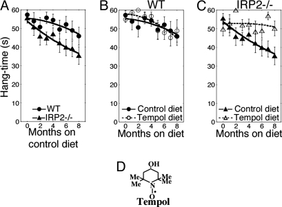
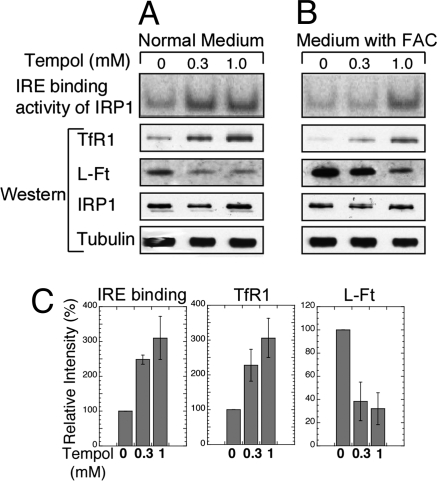
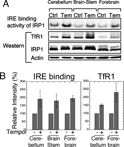
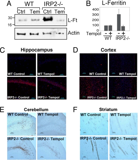
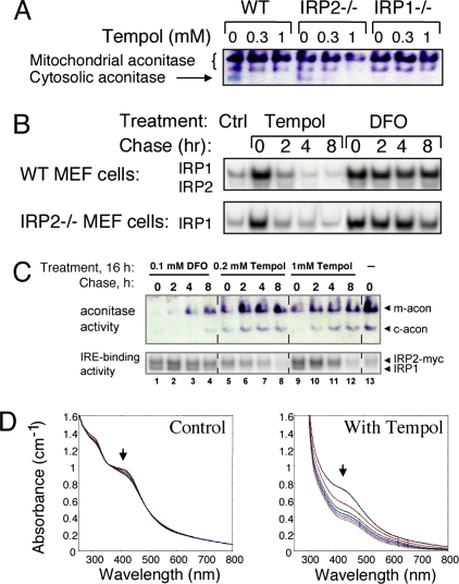
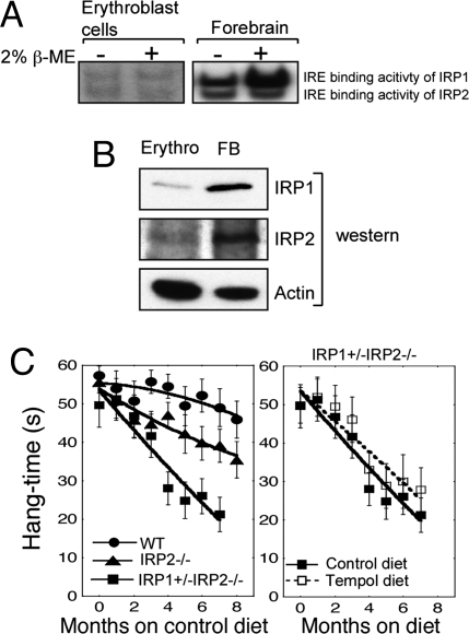
In mammals, two homologous cytosolic regulatory proteins, iron regulatory protein 1 (also known as IRP1 and Aco1) and iron regulatory protein 2 (also known as IRP2 and Ireb2), sense cytosolic iron levels and posttranscriptionally regulate iron metabolism genes, including transferrin receptor 1 (TfR1) and ferritin H and L subunits, by binding to iron-responsive elements (IREs) within target transcripts. Mice that lack IRP2 develop microcytic anemia and neurodegeneration associated with functional cellular iron depletion caused by low TfR1 and high ferritin expression. IRP1 knockout (IRP1(-/-)) animals do not significantly misregulate iron metabolism, partly because IRP1 is an iron-sulfur protein that functions mainly as a cytosolic aconitase in mammalian tissues and IRP2 activity increases to compensate for loss of the IRE binding form of IRP1. The neurodegenerative disease of IRP2(-/-) animals progresses slowly as the animals age. In this study, we fed IRP2(-/-) mice a diet supplemented with a stable nitroxide, Tempol, and showed that the progression of neuromuscular impairment was markedly attenuated. In cell lines derived from IRP2(-/-) animals, and in the cerebellum, brainstem, and forebrain of animals maintained on the Tempol diet, IRP1 was converted from a cytosolic aconitase to an IRE binding protein that stabilized the TfR1 transcript and repressed ferritin synthesis. We suggest that Tempol protected IRP2(-/-) mice by disassembling the cytosolic iron-sulfur cluster of IRP1 and activating IRE binding activity, which stabilized the TfR1 transcript, repressed ferritin synthesis, and partially restored normal cellular iron homeostasis in the brain.


Conflict of interest statement

The authors declare no conflict of interest.

Figures

Fig. 1.
Fig. 1.
Dietary Tempol supplementation prevents progressive neuromuscular deterioration in IRP2−/− mice. (A) Hang test studies on cohorts of WILD-TYPE and IRP2−/− animals revealed progressive neuromuscular compromise of IRP2−/− animals (n = 22) compared with wild type (n = 22) for animals maintained on the control diet (see Materials and Methods). (B) Hang test results from wild-type animals on a control (n = 22) or Tempol-supplemented diet (n = 22). (C) IRP2−/− animals on a diet supplemented with Tempol (n = 22) markedly improved their performance on the hang test compared with the animals on the control diet (n = 22). (D) The structure of Tempol is shown. Error bars represent standard error of the mean. The curves were drawn by using the polynomial curve fit of the KaleidaGraph program (Synergy).
Fig. 2.
Fig. 2.
Tempol supplementation activated the IRE binding activity of IRP1, increased TfR1 levels, and decreased ferritin expression in mouse embryonic fibroblasts derived from IRP2−/− animals. (A) IRP2−/− embryonic fibroblasts were maintained in cultures supplemented with 0, 0.3, or 1.0 mM Tempol for 16 h. In cells that lacked IRP2, all IRE binding activity in the first row was attributable to IRP1 activation. Western blots revealed that TfR1 levels increased, and ferritin levels decreased in Tempol treated cells, whereas IRP1 and tubulin levels (loading control) did not significantly change. (B) IRP2−/− embryonic fibroblasts were maintained in cultures supplemented with 300 μM ferric ammonium citrate (FAC) and 0, 0.3, or 1.0 mM Tempol for 16 h. Increased IRE binding activity of IRP1 correlated with increased TfR1 levels and decreased ferritin levels. (C) IRE binding activities of IRP1 and TfR1 and L-Ft protein levels in the absence and presence of two different concentrations of Tempol without added FAC were quantified in comparison with the intensity of the control lanes, represented as 100%. Error bars represent the standard deviation calculated from the results of two different sets of experiments.
Fig. 3.
Fig. 3.
IRE binding activity of IRP1 was increased in the brains of 11-mo-old IRP2−/− animals maintained for 10 mo on a Tempol-supplemented diet, resulting in increased expression of TfR1. (A) IRP2−/− animals on control (Ctrl) or Tempol (Tem) diets were killed, and lysates from the cerebellum, forebrain, and brain stem were analyzed by gel-shift assay and Western blot. IRE binding activity of IRP1 was increased by Tempol supplementation and TfR1 levels increased concomitantly, whereas total IRP1 and actin levels (loading control) did not change significantly after Tempol treatment. (B) IRE binding activities of IRP1 and TfR1 protein levels in different brain tissues from Tempol-supplemented mice were quantified relative to the intensity of the control mouse bands, represented as 100%. Error bars represent the standard deviation calculated from the results of two different sets of animals.
Fig. 4.
Fig. 4.
Increased IRE binding activity induced by Tempol treatment leads to decreased ferritin expression in brain and reduced ferric iron accumulation in white matter. (A) Ferritin levels detected in Western blot of cerebellar lysates of wild-type animals on a control (Ctrl) (lane 1) or Tempol (Temp) diet (lane 2) compared with IRP2−/− animals on a control (lane 3) or Tempol diet (lane 4) indicated that increased cerebellar ferritin levels of untreated IRP2−/− animals were markedly reduced by treatment with Tempol. (B) L-Ft protein levels in the cerebellum from IRP2−/− mice on a control diet and Tempol-supplemented wild-type and IRP2−/− mice were quantified relative to the intensity of the wild-type control mouse bands, represented as 100%. Error bars represent the standard deviation calculated from the results of three different sets of animals. (C) Ferritin immunohistochemistry performed on animals on control diets revealed marked increases in ferritin expression in hippocampal neurons in IRP2−/− animals (Lower Left), which decreased with Tempol treatment (Lower Right). (D) Tempol treatment diminished ferritin overexpression in the cortex of IRP2−/− mice. (E) Cerebellar folia from wild-type and IRP2−/− animals were stained with Perls' DAB stain for detection of ferric iron. Ferric-iron staining increased in the white matter of IRP2−/− animals on a control diet compared with wild-type controls, but decreased in IRP2−/− animals on the Tempol diet. (F) Ferric-iron staining was also increased in the striatum of IRP2−/− animals but diminished in IRP2−/− animals on Tempol supplementation.
Fig. 5.
Fig. 5.
Tempol treatment activates IRP1 to bind IREs by converting IRP1 from its cytosolic aconitase form to the IRE binding form. (A) The cytosolic aconitase activity in mouse embryonic fibroblast (MEF) lysates (arrow), as assessed in the in-gel aconitase assay for mouse lysates, was identified by its absence in IRP1−/− cells and was diminished by Tempol treatment of cells, as observed in lanes 2, 3, 5, and 6. (B) MEFs from wild-type or IRP2−/− animals were treated with 1.0 mM Tempol or 0.1 mM DFO for 16 h, after which they were switched to fresh unsupplemented media and assayed at various time points with gel-shift and aconitase gel assays. Recovery of aconitase activity was assessed in wild-type and IRP2−/− cells. (C) Aconitase activity (Upper) and IRE binding activity (Lower) of a HEK cell line engineered to overexpress myc-tagged IRP2 was assessed after 16 h treatments with media alone (lane 1), 0.1 mM DFO (lanes 2–6), 0.2 mM Tempol (lanes 6–9), or 1 mM Tempol (lanes 10–13). (D) Tempol degrades a purified synthetic [4Fe–4S] cluster. Repetitive spectral scanning of ≈60 μM solution of (Bu4N)2[Fe4S4(SEt)4] in acetonitrile maintained under anaerobic conditions over 140 min at 20-min intervals revealed minimal changes in the absorption profile (Left). Repetitive spectral scanning at 20-min intervals of ≈60 μM solution of (Bu4N)2[Fe4S4(SEt)4] to which a 9 mM solution of Tempol in acetonitrile was added demonstrated loss of characteristic absorption of the iron–sulfur cluster at 298 nm and 420 nm (Right).
Fig. 6.
Fig. 6.
Restoration of normal iron homeostasis by Tempol treatment depends on conversion of sufficient amounts of IRP1 to the IRE binding form. (A) Gel-shift studies demonstrate that erythoblast lysates contain little IRP1 that can be converted to the IRE binding form by treatment with 2% 2-mercaptoethanol, whereas forebrain lysates contain large amounts of IRP1 that can be recruited to bind IREs. Twenty micrograms of total protein was loaded in each lane. (B) Lysates from erythroblasts and forebrain indicate that IRP1 and IRP2 levels are low in erythroblasts (Erythro) compared with forebrain (FB). Twenty micrograms of total protein was loaded in each lane. (C) Hang test results of wild-type (n = 22), IRP2−/− (n = 22), and IRP1+/− IRP2−/− (n = 6) animals indicate that IRP1+/− IRP2−/− animals are more symptomatic than IRP2−/− animals [hang test curves of wild-type and IRP2−/− mice shown in Fig. 1 are redisplayed for comparison with IRP1+/− IRP2−/− animals (Left)]. However, Tempol treatment did not protect IRP1+/− IRP2−/− animals significantly (P = 0.559) from progression of neuromuscular compromise (Right). Error bars represent standard error of the mean. The curves were drawn by using the polynomial curve fit of the KaleidaGraph program.

Similar articles

Cited by

References

    1. Rouault TA. The role of iron regulatory proteins in mammalian iron homeostasis and disease. Nat Chem Biol. 2006;2:406–414. - PubMed
    1. Wallander ML, Leibold EA, Eisenstein RS. Molecular control of vertebrate iron homeostasis by iron regulatory proteins. Biochim Biophys Acta. 2006;1763:668–689. - PMC - PubMed
    1. Hentze MW, Muckenthaler MU, Andrews NC. Balancing acts: Molecular control of mammalian iron metabolism. Cell. 2004;117:285–297. - PubMed
    1. Dupuy J, et al. Crystal structure of human iron regulatory protein 1 as cytosolic aconitase. Structure. 2006;14:129–139. - PubMed
    1. Walden WE, et al. Structure of dual function iron regulatory protein 1 complexed with ferritin IRE-RNA. Science. 2006;314:1903–1908. - PubMed

Publication types

MeSH terms