. 2008 Aug 12;105(32):11122-7.
doi: 10.1073/pnas.0805399105. Epub 2008 Aug 6.

Human mitochondrial RNA polymerase primes lagging-strand DNA synthesis in vitro

Affiliations

Human mitochondrial RNA polymerase primes lagging-strand DNA synthesis in vitro

Sjoerd Wanrooij et al. Proc Natl Acad Sci U S A. .

Abstract

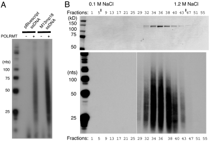
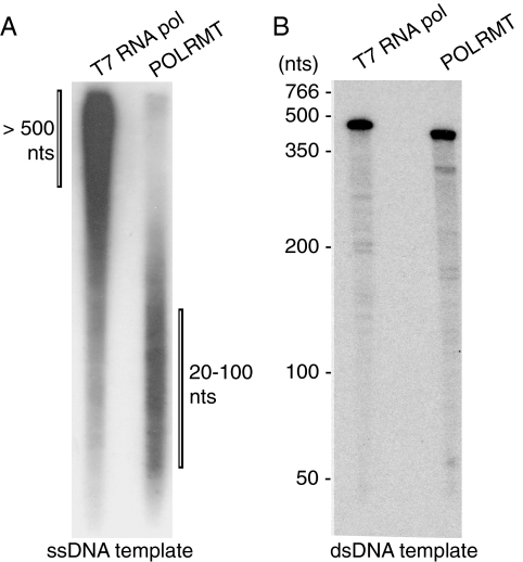
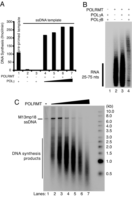
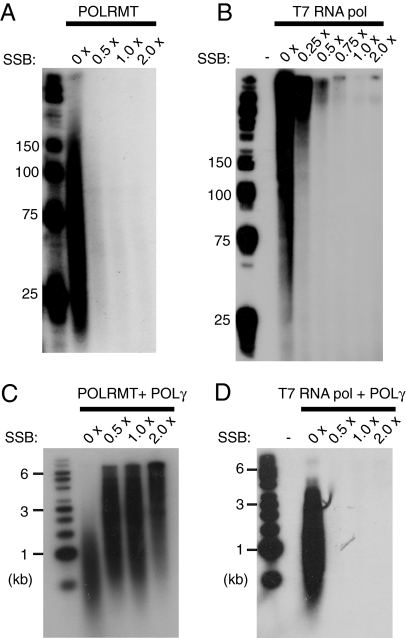
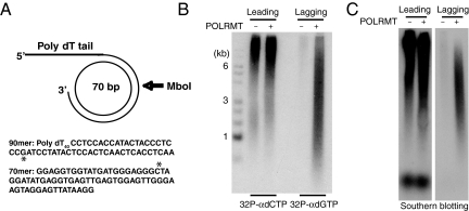
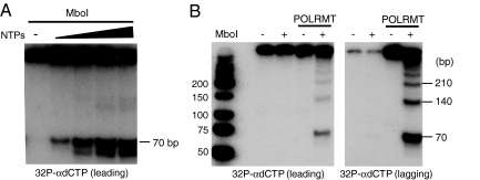
The mitochondrial transcription machinery synthesizes the RNA primers required for initiation of leading-strand DNA synthesis in mammalian mitochondria. RNA primers are also required for initiation of lagging-strand DNA synthesis, but the responsible enzyme has so far remained elusive. Here, we present a series of observations that suggests that mitochondrial RNA polymerase (POLRMT) can act as lagging-strand primase in mammalian cells. POLRMT is highly processive on double-stranded DNA, but synthesizes RNA primers with a length of 25 to 75 nt on a single-stranded template. The short RNA primers synthesized by POLRMT are used by the mitochondrial DNA polymerase gamma to initiate DNA synthesis in vitro. Addition of mitochondrial single-stranded DNA binding protein (mtSSB) reduces overall levels of primer synthesis, but stimulates primer-dependent DNA synthesis. Furthermore, when combined, POLRMT, DNA polymerase gamma, the DNA helicase TWINKLE, and mtSSB are capable of simultaneous leading- and lagging-strand DNA synthesis in vitro. Based on our observations, we suggest that POLRMT is the lagging-strand primase in mammalian mitochondria.


Conflict of interest statement

The authors declare no conflict of interest.

Figures

Fig. 1.
Fig. 1.
POLRMT synthesizes short RNA stretches on ssDNA templates. RNA synthesis by POLRMT was monitored as described in Material and Methods by labeling the RNA. (A) RNA synthesis by POLRMT (500 fmol) was carried out on pBluescript or M13mp18 ssDNA templates and the products were separated on a denaturing polyacrylamide gel (10%). (B) POLRMT peak fractions (10 μl) from purification over MonoS were separated by SDS/PAGE (10%) and revealed with Coomassie Brilliant Blue staining (Upper). The RNA synthesis of the protein fractions (5 μl) in the upper panel was analyzed in the presence of M13mp18 ssDNA (Lower). The beginning and the end of the linear salt gradient are indicated.
Fig. 2.
Fig. 2.
RNA synthesis of POLRMT and T7 RNA polymerase on ssDNA and dsDNA templates. RNA synthesis by POLRMT (500 fmol) or by T7 RNA polymerase (0.8 units) were monitored as described in Material and Methods. (A) RNA products formed on the M13mp18 ssDNA template were separated on a denaturing polyacrylamide gel (10%). (B) RNA products formed by promoter-dependent transcription on a dsDNA template were separated on a denaturing polyacrylamide gel (10%). The positions of molecular size markers are indicated on the left. The length of the T7 RNA polymerase product is 458 nt and the POLRMT product is 396 nt.
Fig. 3.
Fig. 3.
POLRMT primes DNA synthesis by POLγ on ssDNA templates. (A) RNA-primed DNA synthesis on an ssDNA template was monitored in the presence of [α-32P]dCTP for the labeling of leading-strand products. Reactions (20 μl) were incubated for 1 h at 37°C and aliquots were applied to DE81 filter papers for further analysis and scintillation counting as described in Material and Methods. Bar 1, Control experiment using preprimed M13mp18 ssDNA with POLγ (100 fmol); Bar 2, POLRMT (200 fmol); Bar 3, POLγ (100 fmol); Bar 4–7, POLRMT (200 fmol) and increasing amounts of POLγ (50, 100, 500, and 1000 fmol). (B) RNA-primed DNA synthesis on ssDNA was performed in the presence of [α-32P]UTP for labeling of RNA primers. Reactions (25 μl) contained M13mp18 ssDNA (35 fmol) and, when indicated, POLRMT (500 fmol), POLγA (100 fmol), or POLγB (300 fmol). After incubation for 1 h at 37°C, the products were separated on a denaturing polyacrylamide gel (10%) and detected by autoradiography. (C) The RNA-primed DNA synthesis assay was performed in the presence of [α-32P]dCTP. Reactions (20 μl) contained M13mp18 ssDNA (35 fmol), POLγ (100 fmol), and increasing amounts of POLRMT (0, 25, 50, 100, 250, 500, and 1000 fmol). After incubation for 1 h at 37°C the products were separated by electrophoresis on a denaturing agarose gel (0.8%) and detected by autoradiography.
Fig. 4.
Fig. 4.
MtSSB influences POLRMT-dependent primer synthesis. (A) RNA synthesis on ssDNA was performed as described in Material and Methods. Reactions mixtures (25 μl) contained M13mp18 ssDNA (35 fmol), POLRMT (500 fmol), and increasing amounts of mtSSB (0 fmol, 8.4 pmol, 16.8 pmol, 33.6 pmol). Each mtSSB monomer covers approximately 60 nt (33, 34) and the saturation level for each protein concentration was calculated. The saturation level 1 × indicates that the ssDNA molecules should be completely covered with mtSSB. After the incubation for 1 h at 37°C, reaction products were separated by electrophoresis on a denaturing polyacrylamide gel (10%) and detected by autoradiography. (B) Reactions were performed as in (A), but in the presence of T7 RNA polymerase (1 unit). (C) RNA-primed DNA synthesis on ssDNA in the presence of [α-32P]dCTP for labeling of DNA. Reactions (25 μl) contained M13mp18 ssDNA (35 fmol), POLRMT (500 fmol), POLγA (100 fmol), POLγB (300 fmol), and increasing amounts of mtSSB [saturation levels are indicated in (A)]. The reactions were allowed to proceed for 1 h at 37°C and the reaction products were separated by electrophoresis on a denaturing agarose gel (1%) and detected by autoradiography. (D) Reactions were performed as in (C), but in the presence of T7 RNA polymerase (1 unit).
Fig. 5.
Fig. 5.
Lagging-strand DNA synthesis in vitro. (A) Minicircle template for rolling-circle DNA replication. Radioactive dNTPs can be used to preferentially label the synthesis of leading ([α-32P]dCTP) or lagging ([α-32P]dGTP) strands. The single G and C in the templates for lagging and leading strands are indicated with an asterisk. (B and C) Products formed by rolling-circle DNA replication in vitro were analyzed by electrophoresis on an alkaline agarose gel as described in Materials and Methods. The reaction mixtures contained POLγ (200 fmol), TWINKLE (100 fmol), mtSSB (5 pmol), and POLRMT (200 fmol) as indicated. (B) POLRMT supports lagging-strand DNA synthesis. Lane 1, DNA size markers; lanes 2 and 3, leading-strand DNA synthesis labeled with [α-32P]dCTP; lanes 4 and 5, lagging-strand DNA synthesis labeled with [α-32P]dGTP. (C) Southern blot analyses of replication products. In lanes 1 and 2, the oligonucleotide probe is complementary to the leading strand; in lanes 3 and 4, the oligonucleotide probe is complementary to the lagging strand.
Fig. 6.
Fig. 6.
Double-stranded DNA synthesis in vitro. (A) Production of dsDNA depends on NTPs. Rolling-circle DNA replication was performed as described in figure legend 5, but in the presence of varying amounts of NTPs (0 μM, 37.5 μM, 75 μM, 150 μM, and 300 μM). Products were labeled with [α-32P]dCTP, cleaved with MboI, separated by polyacrylamide gel electrophoresis, and detected by autoradiography. (B) Incomplete cleavage by MboI detects long double-stranded DNA products, indicative of rolling-circle DNA replication. The analysis was as in (A), but products labeled with either [α-32P]dGTP (lagging strand) or [α-32P]dCTP (leading strand) were cleaved with subsaturating amounts of MboI.

Similar articles

Cited by

References

    1. Falkenberg M, Larsson NG, Gustafsson CM. DNA replication and transcription in mammalian mitochondria. Annu Rev Biochem. 2007;76:679–699. - PubMed
    1. Robberson DL, Kasamatsu H, Vinograd J. Replication of mitochondrial DNA. Circular replicative intermediates in mouse L cells. Proc Natl Acad Sci USA. 1972;69:737–741. - PMC - PubMed
    1. Tapper DP, Clayton DA. Mechanism of replication of human mitochondrial DNA. Localization of the 5′ ends of nascent daughter strands. J Biol Chem. 1981;256:5109–5115. - PubMed
    1. Kang D, Miyako K, Kai Y, Irie T, Takeshige K. In vivo determination of replication origins of human mitochondrial DNA by ligation-mediated polymerase chain reaction. J Biol Chem. 1997;272:15275–15279. - PubMed
    1. Brown TA, Cecconi C, Tkachuk AN, Bustamante C, Clayton DA. Replication of mitochondrial DNA occurs by strand displacement with alternative light-strand origins, not via a strand-coupled mechanism. Genes Dev. 2005;19:2466–2476. - PMC - PubMed

Publication types

MeSH terms