. 2008 Nov;79(5):954-61.
doi: 10.1095/biolreprod.108.069260. Epub 2008 Aug 6.

Region-specific gene expression profiling: novel evidence for biological heterogeneity of the human amnion

Affiliations

Region-specific gene expression profiling: novel evidence for biological heterogeneity of the human amnion

Yu Mi Han et al. Biol Reprod. 2008 Nov.

Abstract

The amnion plays an important role during pregnancy and parturition. Though referred to as a single structure, this fetal tissue is regionally divided into placental amnion, reflected amnion, and umbilical amnion. Histological differences between placental amnion and reflected amnion led us to hypothesize that the amnion is biologically heterogeneous. The gene expression profiles of placental amnion and reflected amnion were compared in patients at term with no labor (TNL; n = 10) and in labor (TIL; n = 10). Real-time quantitative RT-PCR revealed a higher expression of IL1B mRNA in reflected amnion than in placental amnion in TNL cases but not in TIL cases. Extended screening using microarrays showed differential expression of 17 genes in labor, regardless of the region. Interestingly, 839 genes were differentially expressed between placental amnion and reflected amnion. Pathway analysis identified 19 signaling pathways, such as mitogen-activated protein kinase and transforming growth factor beta pathways, associated with region. Lipopolysaccharide (LPS) treatment of the amnion explants showed more robust activation of mitogen-activated protein kinase 3/1 (extracellular signal-regulated kinase 1/2) in placental amnion of TNL but not in TIL cases. Placental amnion from TNL and TIL cases showed a significant difference in the amplitude of IL1B mRNA induction by LPS. We report that the anatomical region has a substantial impact on the transcriptional program and the biological properties of the amnion. Labor-associated switching to a proinflammatory signature is a feature particular to placental amnion. The novel observations herein strongly suggest that the seemingly homogeneous amnion is biologically heterogeneous and compartmentalized, with implications for the physiology of pregnancy and parturition.


Figures
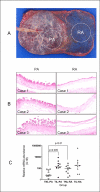
FIG. 1.
FIG. 1.
Differences in the morphological reaction and IL1B mRNA expression between placental amnion (PA) and reflected amnion (RA). A) The image shows representative sampling sites in each placenta, PA and RA. B) Reactive changes in PA epithelial cells induced by meconium are in stark contrast to relatively minimal changes in RA of the same placenta. Three examples are shown. Original magnification ×100. C) Expression patterns of IL1B mRNA in PA and RA of term with no labor (TNL) and term in labor (TIL) groups. There is a significant difference in IL1B mRNA expression between PA of TNL and TIL cases (P < 0.005) indicating that robust change associated with labor at term is more characteristic of PA. IL1B mRNA expression is significantly higher in PA than in RA (P < 0.01) in TNL but not TIL groups.
FIG. 2.
FIG. 2.
The results of transcriptomic analysis of placental amnion (PA) and reflected amnion (RA). A) The principal component (PC) analysis plot shows clear segregation of the data according to the region (PA vs. RA). TNL_PA: gray, TNL_RA: magenta, TIL_PA: blue, TIL_RA: black. B) The volcano plots show significance P values plotted against the fold changes. In this figure the log (base 10) of the FDR-adjusted P values are plotted against the log (base 2) of fold changes: the higher the y-axis, the more significant the gene expression changes. the magnitude of difference between groups, as computed by the fold changes, is larger as the departure from 0 on the x-axis increases. C) A heat map showing clustering of both genes and samples. The genes used in the clustering were filtered unbiasedly using a variance threshold (TNL_PA: gray, TNL_RA: magenta, TIL_PA: blue, TIL_RA: black).
FIG. 3.
FIG. 3.
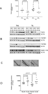
Expression patterns of transforming growth factor beta signaling components, laser microdissection of amnion for BNC2 (basonuclin-2) mRNA expression analysis, and PTGS2 mRNA expression. A) TGFBR1 mRNA expression is significantly higher in placental amnion (PA) than in reflected amnion (RA) in TNL cases (P < 0.01). A similar tendency is also observed in TIL cases. B) Immunoblots for TGFB signaling proteins, TGFB1/TGFB2/TGFB3, TGFBR1, TGFBR2, and SMAD1/SMAD5/ SMAD9. All the blots taken from both TNL and TIL groups show a higher expression of signaling molecules in reflected amnion than in placental amnion. TGFBR1 protein expression is higher in reflected amnion, which is paradoxical to TGFBR1 mRNA. C) Amnion epithelial layer or mesodermal layer were selectively procured for RNA isolation and subsequent qRT-PCR reaction (1: image of amnion before microdissection, 2: image after ablation of mesodermal layer, 3: image after microdissection of amnion epithelial cells). The arrows indicate the amnion epithelial layer. Original magnification ×50. D) The results of qRT-PCR for PTGS2 mRNA show increased expression in both placental amnion (P = 0.054) and reflected amnion (P = 0.001) with labor. PTGS2 mRNA expression is higher in placental amnion compared to reflected amnion in TNL but not TIL cases (P = 0.017).
FIG. 4.
FIG. 4.
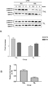
Lipopolysaccharide (LPS)-induced biological response in placental amnion. A) LPS induces phosphorylation of MAPK3/MAPK1 (ERK 1/2). MAPK3/MAPK1 activation is stronger and more sustained in placental amnion (PA) than in reflected amnion (RA) of TNL case but not TIL case. The graph shows the relative intensity of MAPK3 phosphorylation at 4 h after LPS treatment compared to 0 h on densitometry. The intensity of phosphorylated MAPK3 was divided by the intensity of total MAPK3. Each group is composed of three cases. B) The comparison of IL1B mRNA induction in placental amnion of TNL and TIL cases 4 h after LPS treatment. Three cases were evaluated in both TNL and TIL groups. The fold increases were 4.9 and 1.7 in placental amnion of TNL and TIL cases, respectively.

References

    1. Millar LK, Stollberg J, Debuque L, Bryant-Greenwood G.Fetal membrane distention: determination of the intrauterine surface area and distention of the fetal membranes preterm and at term. Am J Obstet Gynecol 2000; 182: 128–134.. - PubMed
    1. Benirschke K, Kaufmann P.Anatomy and pathology of the placental membranes. Pathology of the Human Placenta, 4th ed New York:Springer;2000: 281–334..
    1. Arikat S, Novince RW, Mercer BM, Kumar D, Fox JM, Mansour JM, Moore JJ.Separation of amnion from choriodecidua is an integral event to the rupture of normal term fetal membranes and constitutes a significant component of the work required. Am J Obstet Gynecol 2006; 194: 211–217.. - PubMed
    1. Oyen ML, Calvin SE, Landers DV.Premature rupture of the fetal membranes: is the amnion the major determinant? Am J Obstet Gynecol 2006; 195: 510–515.. - PubMed
    1. Parry S, Strauss JF., IIIPremature rupture of the fetal membranes. N Engl J Med 1998; 338: 663–670.. - PubMed

Publication types