

Biventricular apical thrombi demonstrated by contrast-enhanced cardiac MRI following anteroapical STEMI and unsuccessful reperfusion therapy
- PMID: 18685749
- PMCID: PMC2644367
- DOI: 10.1016/s0828-282x(08)70655-x
Biventricular apical thrombi demonstrated by contrast-enhanced cardiac MRI following anteroapical STEMI and unsuccessful reperfusion therapy
Abstract
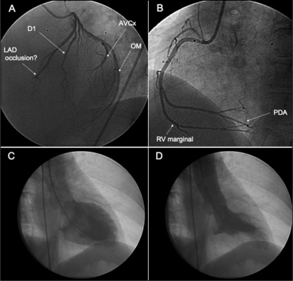
Contrast-enhanced cardiac magnetic resonance imaging can define the territory and extent of myocardial infarction from patterns of late gadolinium enhancement. Following failure to reperfuse with thrombolytic therapy, a case of myocardial infarction is described in which ongoing symptoms and an electrocardiogram change led to a diagnostic dilemma. Cardiac magnetic resonance imaging confirmed an apical infarction, an aneurysm and acute pericarditis. In addition, late gadolinium enhancement unexpectedly revealed the presence of biventricular apical thrombi. The prevalence of cardiac thrombi and pulmonary emboli may be greater than generally appreciated.
L’imagerie par résonance magnétique cardiaque à contraste rehaussé permet de définir le territoire et l’ampleur de l’infarctus du myocarde à partir de l’image de phase tardive rehaussée par le gadolinium. Après l’échec de la reperfusion par thrombolytiques, on décrit un cas d’infarctus du myocarde au cours duquel les symptômes persistants et les changements à l’ÉCG ont mené à un dilemme diagnostique. L’imagerie par résonance magnétique cardiaque a confirmé un infarctus apical, un anévrisme et une péricardite aiguë. De plus, la phase tardive du rehaussement au gadolinium a révélé de manière imprévue la présence de thrombi apicaux biventriculaires. La prévalence des thrombi cardiaques et des embolies pulmonaires pourrait se révéler plus forte qu’on ne l’a généralement cru.
Figures


References
-
- Teraoka K, Kiuchi S, Takada N, Hirano M, Yamashina A. No delayed enhancement on contrast magnetic resonance imaging with Takotsubo cardiomyopathy. Circulation. 2005;111:e261–2. - PubMed
-
- Cury RC, Abbara S, Sandoval LJ, Houser S, Brady TJ, Palacios IF. Visualization of endomyocardial fibrosis by delayed-enhancement magnetic resonance imaging. Circulation. 2005;111:e115–7. - PubMed
-
- Barkhausen J, Hunold P, Eggebrecht H, et al. Detection and characterization of intracardiac thrombi on MR imaging. AJR Am J Roentgenol. 2002;179:1539–44. - PubMed
-
- Mollet NR, Dymarkowski S, Volders W, et al. Visualization of ventricular thrombi with contrast-enhanced magnetic resonance imaging in patients with ischemic heart disease. Circulation. 2002;106:2873–6. - PubMed
-
- Ogren M, Bergqvist D, Eriksson H, Lindblad B, Sternby NH. Prevalence and risk of pulmonary embolism in patients with intracardiac thrombosis: A population-based study of 23 796 consecutive autopsies. Eur Heart J. 2005;26:1108–14. - PubMed
Publication types
MeSH terms
Substances
LinkOut - more resources
Full Text Sources
Medical
