. 2008 Aug 7:7:65.
doi: 10.1186/1476-4598-7-65.

The parafibromin tumor suppressor protein interacts with actin-binding proteins actinin-2 and actinin-3

Affiliations

The parafibromin tumor suppressor protein interacts with actin-binding proteins actinin-2 and actinin-3

Sunita K Agarwal et al. Mol Cancer. .

Abstract

Background: Germline and somatic inactivating mutations in the HRPT2 gene occur in the inherited hyperparathyroidism-jaw tumor syndrome, in some cases of parathyroid cancer and in some cases of familial hyperparathyroidism. HRPT2 encodes parafibromin. To identify parafibromin interacting proteins we used the yeast two-hybrid system for screening a heart cDNA library with parafibromin as the bait.

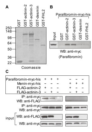
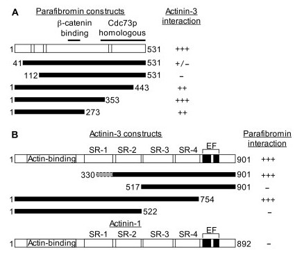
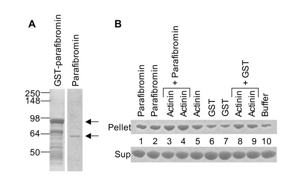
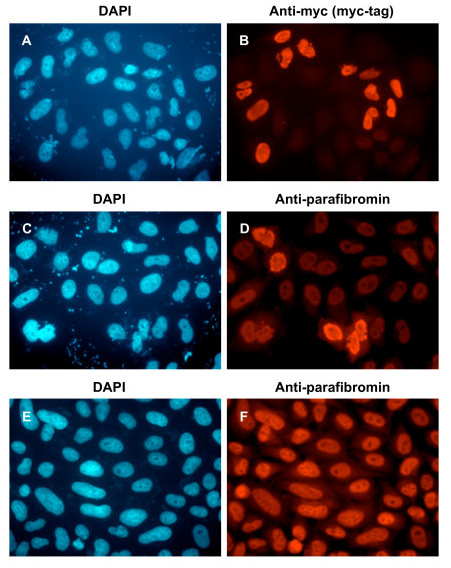
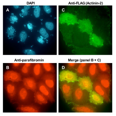
Results: Fourteen parafibromin interaction positive preys representing 10 independent clones encoding actinin-2 were isolated. Parafibromin interacted with muscle alpha-actinins (actinin-2 and actinin-3), but not with non-muscle alpha-actinins (actinin-1 and actinin-4). The parafibromin-actinin interaction was verified by yeast two-hybrid, GST pull-down, and co-immunoprecipitation. Yeast two-hybrid analysis revealed that the N-terminal region of parafibromin interacted with actinins. In actin sedimentation assays parafibromin did not dissociate skeletal muscle actinins from actin filaments, but interestingly, parafibromin could also bundle/cross-link actin filaments. Parafibromin was predominantly nuclear in undifferentiated proliferating myoblasts (C2C12 cells), but in differentiated C2C12 myotubes parafibromin co-localized with actinins in the cytoplasmic compartment.

Conclusion: These data support a possible contribution of parafibromin outside the nucleus through its interaction with actinins and actin bundling/cross-linking. These data also suggest that actinins (and actin) participate in sequestering parafibromin in the cytoplasmic compartment.


Figures

Figure 1
Figure 1
Verification of yeast two-hybrid interaction of parafibromin with actinin. (A) GST or GST-fusion proteins used for in vitro binding assay were analyzed by SDS-PAGE and stained with Coomassie Blue. On the left are shown molecular weights of the protein standards in kilodaltons. (B) GST pull-down assay: GST or GST-fusion proteins coupled to glutathione sepharose beads were incubated with whole cell protein extracts from HEK293 cells transfected with plasmid expressing parafibromin-myc-his. The beads were washed thoroughly and the bound parafibromin was detected by western blotting (WB) with an anti-myc antibody. Input corresponds to 1/40th of the amount of protein extracts used for the pull-down assay. (C) Co-immunoprecipitation assay: Whole cell protein extracts from HEK293 cells transfected with plasmids expressing parafibromin-myc-his or menin-myc-his alone or together with FLAG-actinin-2 or FLAG-actinin-3 were immunoprecipitated with a rabbit anti-myc antibody. The immunoprecipitates were analyzed by western blotting (WB) with a mouse anti-myc (to detect parafibromin) or anti-FLAG (to detect actinin) antibody. Input panels show portions of protein extracts corresponding to 1/40th of the amount used for each immunoprecipitation (IP), probed with anti-myc or anti-FLAG antibody.
Figure 2
Figure 2
Parafibromin and actinin interacting regions. (A) Schematic of the 531 amino acid (aa) parafibromin protein. Striped regions are the 3 nuclear localization signals (aa 76–92, 125–139, and 393–409). The 2 black lines on top show the β-catenin interacting region (aa 218–263), and the Cdc73p homologous region (aa 343–531). Below are shown regions of parafibromin fused to the Gal4DBD that were tested for interaction with Gal4AD-actinin-3 by yeast two-hybrid assays as described in the footnote of Table 2. (B) Schematic of actinin-3 protein (901 amino acids). Following the N-terminal actin-binding region are 4 striped areas corresponding to the 4 spectrin repeats: SR-1 (aa 288–398), SR-2 (aa 408–512), SR-3 (aa 523–634), and SR-4 (aa 642–747). The two black areas near the C-terminus are the EF-hand domains (aa 764–793, and 800–828). Below are shown regions of actinin-3 fused to the Gal4AD that were tested for interaction with Gal4DBD-parafibromin by yeast two-hybrid assays as described in the footnote of Table 2. The dashed grey shaded area (aa 330–397) in the first actinin-3 construct corresponds to the location of the truncated N-terminus (aa 323, 327, 329, 333, 340, 360, 370, 377, 381 and 390) of the 10 independent actinin-2 clones, in-frame with the Gal4AD, isolated by yeast two-hybrid library screening. Also shown is the schematic of actinin-1 protein (892 amino acids) representing the two non-muscle actinins (actinin-1 and actinin-4) that do not interact with parafibromin. They show over 75% amino acid sequence identity with the muscle actinins (actinin-2 and actinin-3). Their domain composition and domain location is similar to the muscle actinins. However, the muscle actinins possess amino acid substitutions (not shown) in the EF hand domains that prevent calcium binding and render them insensitive to calcium. Calcium binding reduces the affinity of non-muscle actinins for F-actin [35]. Interaction is indicated by β-galactosidase activity level. -, +, ++, or +++ indicate no, low, medium or high levels of β-galactosidase activity, respectively. Activity level intermediate between 2 categories is indicated by a slash separating the 2 categories.
Figure 3
Figure 3
Actin bundling/cross-linking by actinin and parafibromin. (A) GST-parafibromin, and parafibromin separated from GST after thrombin cleavage was analyzed by SDS-PAGE and stained with Coomassie Blue. On the left are shown molecular weights of the protein standards in kilodaltons. Arrows show the bands corresponding to GST-parafibromin (upper arrow for the panel on the left) and parafibromin (lower arrow for the panel on the right). (B) G-actin was polymerized to F-actin. F-actin was incubated with rabbit skeletal muscle actinin, parafibromin (2 different preps) or GST (2 different preps) alone, or together as indicated, and centrifuged at 10,000 g for 15 minutes to sediment higher order structures of F-actin (bundles and/or cross-linked networks). The F-actin in the supernate (not bundled/cross-linked) and pellet (F-actin bundles/networks) fraction was analyzed by SDS-PAGE and stained with Coomassie Blue. Lane 10 (buffer) contains F-actin that was sedimented without any added GST, actinin or parafibromin, and therefore corresponds to the background level of F-actin bundles/networks in the pellet (similar background amount was also seen in the lane 6 and 7 marked GST). Increased actin, above background level, in the pellet fraction (lane 1, 2, 3, 4, 5, 8 and 9) indicates F-actin bundling and/or cross-linking.
Figure 4
Figure 4
Specificity of anti-parafibromin antibody (GRAPE) by immunofluorescence. HeLa cells transfected with plasmid expressing parafibromin-myc-his (A, B, C, D) or untransfected (E, F) were analyzed by immunofluorescence with primary antibodies that recognize the myc-tag (B) or parafibromin (D, F), and secondary antibody conjugated to Texas Red, followed by DAPI staining (A, C, E). Magnification, 60×.
Figure 5
Figure 5
Location of endogenous parafibromin and actinin in C2C12 myoblasts or C2C12 differentiated into myotubes. C2C12 cells were analyzed by immunofluorescence with anti-parafibromin (GRAPE) antibody and anti-actinin antibody before (A, B, C) or after differentiation (D, E, F), and secondary antibody conjugated to Texas Red (to detect parafibromin) or fluorescein isothiocyanate (to detect actinin), followed by DAPI staining (A, D). Note that in panel E, along with myotubes there are some undifferentiated myoblasts that retain nuclear parafibromin staining (arrows). Magnification, 60× (A, B, C), or 10× (D, E, F).
Figure 6
Figure 6
Location of endogenous parafibromin in HEK293 cells transfected with actinin. HEK293 cells transfected with plasmid expressing FLAG-actinin-2 were analyzed by immunofluorescence with primary antibodies that recognize parafibromin (GRAPE) or the FLAG-tag (actinin-2), and secondary antibody conjugated to Texas Red (B, to detect endogenous parafibromin) or fluorescein isothiocyanate (C, to detect transfected actinin-2), followed by DAPI staining (A). Note that in untransfected cells parafibromin is nuclear but in the 3 cells over-expressing FLAG-actinin-2, parafibromin is in the cytoplasm. Merged Texas Red (parafibromin) and fluorescein isothiocyanate (actinin-2) signal is shown in panel D. Magnification, 60×.

References

    1. Carpten JD, Robbins CM, Villablanca A, Forsberg L, Presciuttini S, Bailey-Wilson J, Simonds WF, Gillanders EM, Kennedy AM, Chen JD, Agarwal SK, Sood R, Jones MP, Moses TY, Haven C, Petillo D, Leotlela PD, Harding B, Cameron D, Pannett AA, Höög A, Heath H, 3rd, James-Newton LA, Robinson B, Zarbo RJ, Cavaco BM, Wassif W, Perrier ND, Rosen IB, Kristoffersson U, Turnpenny PD, Farnebo LO, Besser GM, Jackson CE, Morreau H, Trent JM, Thakker RV, Marx SJ, Teh BT, Larsson C, Hobbs MR. HRPT2, encoding parafibromin, is mutated in hyperparathyroidism-jaw tumor syndrome. Nat Genet. 2002;32:676–680. doi: 10.1038/ng1048. - DOI - PubMed
    1. Jackson CE, Norum RA, Boyd SB, Talpos GB, Wilson SD, Taggart RT, Mallette LE. Hereditary hyperparathyroidism and multiple ossifying jaw fibromas: a clinically and genetically distinct syndrome. Surgery. 1990;108:1006–1012. - PubMed
    1. Simonds WF, James-Newton LA, Agarwal SK, Yang B, Skarulis MC, Hendy GN, Marx SJ. Familial isolated hyperparathyroidism: clinical and genetic characteristics of 36 kindreds. Medicine. 2002;81:1–26. doi: 10.1097/00005792-200201000-00001. - DOI - PubMed
    1. Villablanca A, Calender A, Forsberg L, Höög A, Cheng JD, Petillo D, Bauters C, Kahnoski K, Ebeling T, Salmela P, Richardson AL, Delbridge L, Meyrier A, Proye C, Carpten JD, Teh BT, Robinson BG, Larsson C. Germline and de novo mutations in the HRPT2 tumour suppressor gene in familial isolated hyperparathyroidism (FIHP) J Med Genet. 2004;41:e32. doi: 10.1136/jmg.2003.012369. - DOI - PMC - PubMed
    1. Shattuck TM, Välimäki S, Obara T, Gaz RD, Clark OH, Shoback D, Wierman ME, Tojo K, Robbins CM, Carpten JD, Farnebo LO, Larsson C, Arnold A. Somatic and germ-line mutations of the HRPT2 gene in sporadic parathyroid carcinoma. N Engl J Med. 2003;349:1722–1729. doi: 10.1056/NEJMoa031237. - DOI - PubMed

Publication types

Associated data