. 2008 Aug 7:5:73.
doi: 10.1186/1742-4690-5-73.

Nuclear import of Avian Sarcoma Virus integrase is facilitated by host cell factors

Affiliations

Nuclear import of Avian Sarcoma Virus integrase is facilitated by host cell factors

Mark D Andrake et al. Retrovirology. .

Abstract

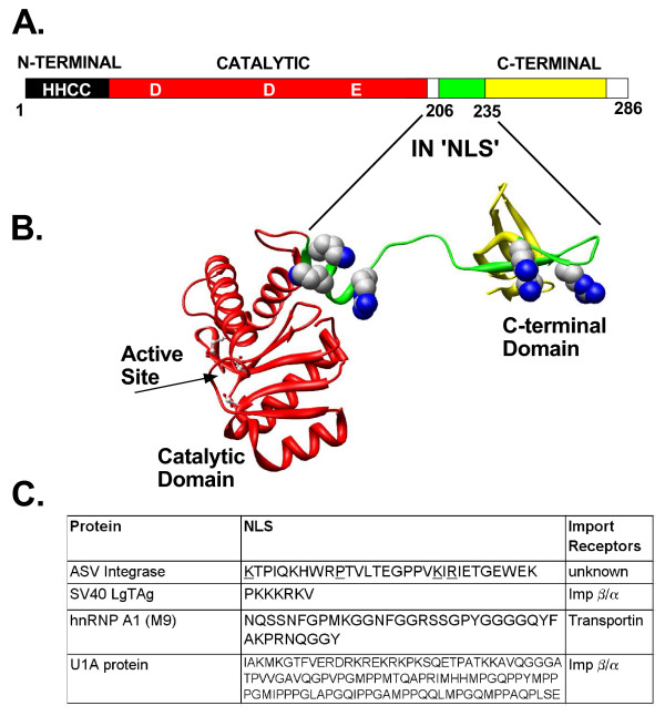
Background: Integration of retroviral DNA into the host cell genome is an obligatory step in the virus life cycle. In previous reports we identified a sequence (amino acids 201-236) in the linker region between the catalytic core and C-terminal domains of the avian sarcoma virus (ASV) integrase protein that functions as a transferable nuclear localization signal (NLS) in mammalian cells. The sequence is distinct from all known NLSs but, like many, contains basic residues that are essential for activity.

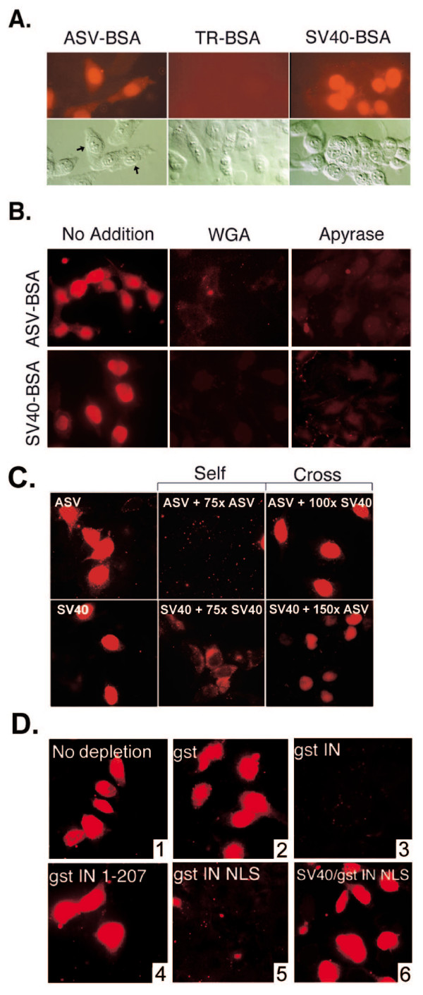
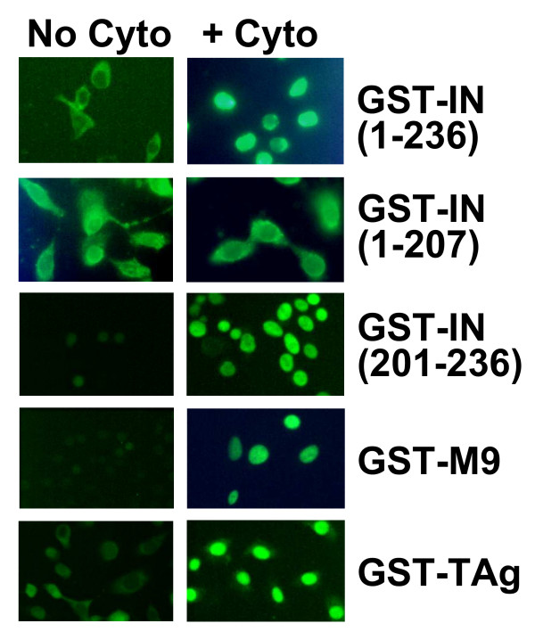
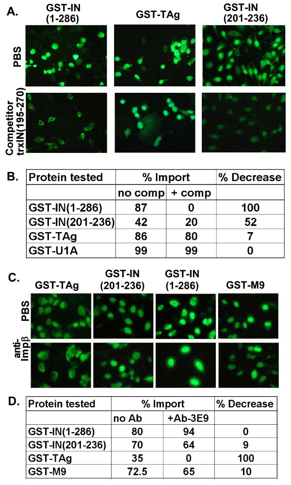
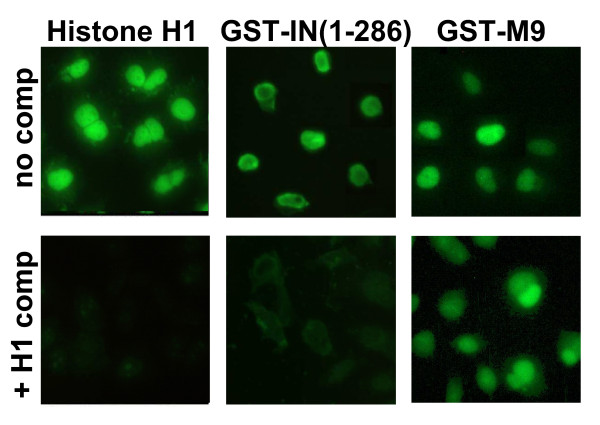
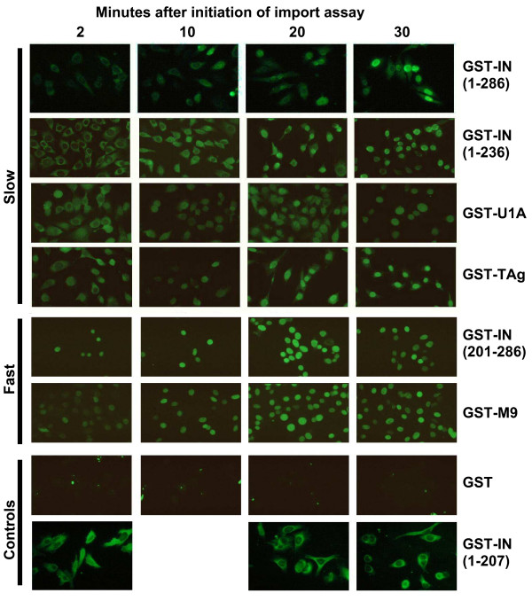
Results: Our present studies with digitonin-permeabilized HeLa cells show that nuclear import mediated by the NLS of ASV integrase is an active, saturable, and ATP-dependent process. As expected for transport through nuclear pore complexes, import is blocked by treatment of cells with wheat germ agglutinin. We also show that import of ASV integrase requires soluble cellular factors but does not depend on binding the classical adapter Importin-alpha. Results from competition studies indicate that ASV integrase relies on one or more of the soluble components that mediate transport of the linker histone H1.

Conclusion: These results are consistent with a role for ASV integrase and cytoplasmic cellular factors in the nuclear import of its viral DNA substrate, and lay the foundation for identification of host cell components that mediate this reaction.


Figures

Figure 1
Figure 1
The ASV IN NLS and three well characterized NLSs. A. Linear map of ASV IN showing the location of NLS sequence. The 286 amino acid IN protein is composed of three domains. The N-terminal, Zn-binding (HHCC) domain (dark) and the central catalytic core domain (red) with the locations of the active site residues (D, D, E) are indicated. The nuclear localization signal, amino acids 206–235 (green), extends from a linker region and into the C-terminal domain (yellow). B. A 3-D structural ribbon model of the catalytic core and C-terminal domains of ASV IN [58] with the with basic residues of the NLS shown in space filling representation. Active site residues in the core domain are shown in ball and stick representation. C. Comparison of the sequences of the ASV IN NLS with three well-characterized NLSs used in the studies reported herein. Residues underlined in the ASV IN NLS have been shown to be required for function.
Figure 2
Figure 2
Nuclear import of ASV-BSA and SV40-BSA substrates; import of ASV-BSA does not require the Impα-Impβ pathway. A. Digitonin-permeabilized HeLa cells were incubated in the presence of complete transport mixture containing the ASV-BSA conjugate, the SV40-BSA conjugate, or Texas red-labeled BSA (TR-BSA). Top panels: Visualization of Texas red conjugates by fluorescence microscopy. Bottom panels: Differential interference contrast (DIC) microscopy of the same field to show preservation of cell integrity. B. Digitonin permeabilized HeLa cells were untreated (no addition), treated with 50 μg/ml wheat germ agglutinin (WGA), or 50 units/ml apyrase (Apyrase) prior to incubation with complete transport mixture containing either the ASV-BSA or the SV40-BSA import substrates. C. Free NLS peptides were added to the import reactions in molar excess of the import substrates as indicated. "Self" signifies competition with the homologous peptides; "Cross" indicates competition for ASV-BSA import by excess SV40TAg NLS peptide or competition for SV40-BSA import by excess ASV NLS peptide. The left column panels show import in the absence of competitor peptides. D. Depletion of ASV-BSA import factor(s) from cytosolic extracts. All assays included Texas-Red labeled ASV-BSA except that shown in the lower left hand corner (panel 4) which included Texas-Red labeled SV40-BSA. Cytosol was either not treated (1; no depletion) or pretreated with glutathione-beads that bound GST alone (2) or fusion proteins of GST plus IN(1–207) which lacks the IN NLS (3), full-length IN(1–286) (5), or a fragment of IN(201–236) that contains the IN NLS (panels 4 and 6).
Figure 3
Figure 3
Nuclear import of GST-NLS substrates in digitonin-permeabilized HeLa cells. GST-NLS fusion proteins were incubated in digitonin permeabilized HeLa cells for 30 min at 37°C prior to fixation with paraformaldehyde and staining with fluorescent antibody against GST. Left column panels are import without added cytosol and right column panels with added HeLa cytosol extracts.
Figure 4
Figure 4
ASV IN NLS import does not compete for import factors required for SV40-TAg and U1A nuclear accumulation. A. Digitonin permeabilized HeLa cells were either treated with buffer (PBS – top row), or with a molar excess of the competitor protein trxIN(195–270) (bottom row). GST-IN(1–286) and GST-TAg had a 15-fold excess of competitor while GST-IN-NLS(201–236) had a 30-fold molar excess. Import assays were performed as shown in Fig. 3 and staining was done with fluorescent antibody against GST. B. Quantitative analysis of nuclear import of various GST fusion proteins with (+ comp) and without (no comp) competitor. More than 100 cells were counted for each experimental condition and the percentage of cells that had a mostly nuclear staining for the fusion protein was calculated. The percent decrease in the presence of the competitor is shown in the column on the right. The lower value for import of GST-IN (201–236) compared to GST-IN (1–286) reflects the fact that a larger percentage of cells had whole cell staining (in which nuclear import could not be assessed) or nuclear exclusion. C. Digitonin permeabilized HeLa cells were either treated with buffer (PBS – top row), or with a 50 ug/ml antibody 3E9 against Impβ (bottom row)during the import reaction. D. Quantitative analysis of nuclear import of various GST fusion proteins with (+ Ab3E9) and without (no Ab) antibody 3E9. More than 100 cells were counted for each experimental condition and the percentage of cells that had a mostly nuclear staining for the fusion protein was calculated. The percent decrease in the presence of the antibody is shown in the column on the right.
Figure 5
Figure 5
ASV IN mediated import is inhibited by excess histone H1. The import of labeled histone H1, (GST-IN(1–286), and the Impβ binding domain fused to GFP (IBB-GFP) was examined in the absence (top) and presence (bottom) of excess unlabeled histone H1. Incubations were for 30 min and all exposure times were equivalent.
Figure 6
Figure 6
Kinetics of ASV-NLS mediated import. GST-NLS fusion proteins were incubated in digitonin permeabilized HeLa cells for various times (labeled above each column) at 37°C prior to fixation with paraformaldehyde and staining with fluorescent antibody against GST. The fusion protein used in each row is labeled at the right and the properties described in the text. Fusion proteins that are imported with slower kinetics are grouped at the top (rows 1–4), and those with faster kinetics in the middle (rows 5 and 6). Control fusion proteins that are not imported into the nucleus are in rows 7 and 8.

References

    1. Fassati A. HIV infection of non-dividing cells: a divisive problem. Retrovirology. 2006;3:74. - PMC - PubMed
    1. Suzuki Y, Craigie R. The road to chromatin - nuclear entry of retroviruses. Nat Rev Microbiol. 2007;5:187–196. - PubMed
    1. Hatziioannou T, Goff SP. Infection of nondividing cells by Rous sarcoma virus. J Virol. 2001;75:9526–9531. - PMC - PubMed
    1. Katz RA, Greger JG, Darby K, Boimel P, Rall GF, Skalka AM. Transduction of interphase cells by avian sarcoma virus. J Virol. 2002;76:5422–5434. - PMC - PubMed
    1. Greger JG, Katz RA, Taganov K, Rall GF, Skalka AM. Transduction of terminally differentiated neurons by avian sarcoma virus. J Virol. 2004;78:4902–4906. - PMC - PubMed

Publication types

MeSH terms