Comparative Study
. 2008 Oct;213(2):439-47.
doi: 10.1016/j.expneurol.2008.07.009. Epub 2008 Jul 18.

Effects of hyperglycemia on rat cavernous nerve axons: a functional and ultrastructural study

Affiliations
Comparative Study

Effects of hyperglycemia on rat cavernous nerve axons: a functional and ultrastructural study

Elena G Zotova et al. Exp Neurol. 2008 Oct.

Abstract

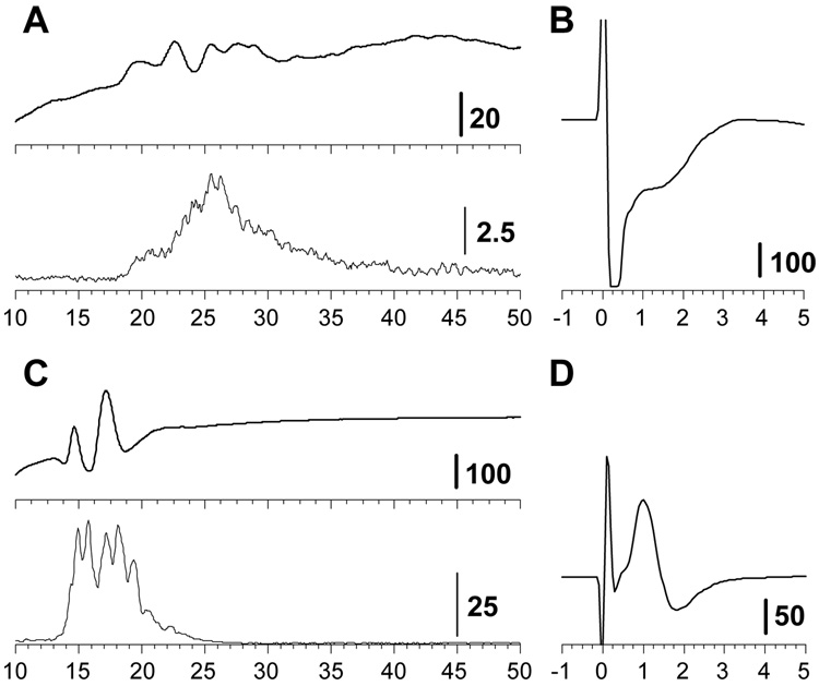
The present study explored parallel changes in the physiology and structure of myelinated (Adelta) and unmyelinated (C) small diameter axons in the cavernous nerve of rats associated with streptozotocin-induced hyperglycemia. Damage to these axons is thought to play a key role in diabetic autonomic neuropathy and erectile dysfunction, but their pathophysiology has been poorly studied. Velocities in slow conducting fibers were measured by applying multiple unit procedures; histopathology was evaluated with both light and electron microscopy. To our knowledge, these are the initial studies of slow nerve conduction velocities in the distal segments of the cavernous nerve. We report that hyperglycemia is associated with a substantial reduction in the amplitude of the slow conducting response, as well as a slowing of velocities within this very slow range (< 2.5 m/s). Even with prolonged hyperglycemia (> 4 months), histopathological abnormalities were mild and limited to the distal segments of the cavernous nerve. Structural findings included dystrophic changes in nerve terminals, abnormal accumulations of glycogen granules in unmyelinated and preterminal axons, and necrosis of scattered smooth muscle fibers. The onset of slowing of velocity in the distal cavernous nerve occurred subsequent to slowing in somatic nerves in the same rats. The functional changes in the cavernous nerve anticipated and exceeded the axonal degeneration detected by morphology. The physiologic techniques outlined in these studies are feasible in most electrophysiologic laboratories and could substantially enhance our sensitivity to the onset and progression of small fiber diabetic neuropathy.


Figures

Figure 1
Figure 1. Conditions of the recording of cavernous nerve activity
A. Schematic diagram illustrates the sites of recording and stimulation for the cavernous nerve and DNP. MPG-major pelvic ganglia, R1-recording site for cavernous nerve activity, R2-recording site for proximal DNP, R3-recording site used for calculation NCV of the distal segment of DNP between R2 and R3, ** - indicates position of stimulation. B. Responses of cavernous nerve are presented as evoked potentials (EP) and multi-unit activity (MUA) at two intensities of stimulation. Open arrows point out initial response of small diameter myelinated fibers (Aδ). Vertical dash lines separate the ranges of NCV. Calibration is given in microvolts, and time scale in milliseconds.
Figure 2
Figure 2. Comparison of the changes in the cavernous nerve activity in the control group and in hyperglycemic rats with diverse severity of neuropathy (moderate and severe)
A. indicates total activity above the baseline averaged across animals characterized as control, moderate or severe neuropathy based on caudal nerve velocity (mean, SEM), B. represents distribution of cavernous nerve activity within various ranges of NCV as a percent of total MUA and averaged across animals for each of these groups.
Figure 3
Figure 3. Changes in the cavernous nerve response observed after different duration of HG
A. MUA responses of the cavernous nerve in control rat and after 2 and 4 months of HG, the last one - at the greater severity of neuropathy. B. Bar graphs on the right illustrate the alterations in responses evaluated by percent of total MUA in various ranges of NCV (marked at the bottom of each graph): disappearance of response in the range of 2–2.5 m/sec and continuous shift of the maximum in response toward the slower NCV. Vertical dash lines - borders of NCV ranges. Calibrations are given in mV; the time scale (in milliseconds) is adapted to fit NCV borders for the activity recorded over different distances.
Figure 4
Figure 4
Comparison of the response in cavernous nerve (A) and possible muscle-twitch related artifact (C) with simultaneous recording evoked activity at DNP (B and D respectively). Calibration is given in µV, and time scale in milliseconds.
Figure 5
Figure 5
Electron microscopy showing: A. A stalk in the crus of a 10-month diabetic. The club-like ultraterminal contains a cluster of normal-appearing smooth muscle cells. (X 3,850). B. Desmosome-like attachment between two smooth muscle fibers. The outer intracellular attachment plaque and the intercellular electron-dense leaflet material within the cleft are visible in the insert. (X 25,000, insert X 125,000). C. Lipid droplets in fibroblasts with adjacent extracellular collagen bundles. (X 3,000). D. Cholesterol clefts in endothelial cells of a helical arteriole. (X 1,950).
Figure 6
Figure 6
A. An electron micrograph of a degenerating smooth muscle cell (sm) with a dystrophic axon preterminal (arrow, in C at the higher magnification, X 62,000) containing dense bodies and degenerating mitochondria. (X 11,500). B. Dystrophic axon preterminal in a six-month diabetic rat containing a few synaptic vesicles, glycogen granules, and a membranous body. (X 56,000).

References

    1. Arezzo JC, Vaughan HG, Jr, Kraut MA, Steinschneider M, Legatt AD. Intracranial generators of event related potentials in the monkey. In: Cracco RQ, Wollner BI, editors. Frontiers of Clinical Neuroscience, Vol. 3 Evoked Potentials. New York: Alan R. Liss, Inc.; 1986. pp. 174–189.
    1. Arezzo JC, Zotova E. Electrophysiologic measures of diabetic neuropathy: mechanism and meaning. In: Tomlinson D, editor. Neurobiology of Diabetic Neuropathy. Amsterdam: Academic Press; 2002. pp. 230–255. - PubMed
    1. Boulton AJ, Vinik AI, Arezzo JC, Bril V, Feldman EL, Freeman R, Malik RA, Maser RE, Sosenko JM, Ziegler D. American Diabetes Association. Diabetic neuropathies: a statement by the American Diabetes Association. Diabetes Care. 2005;28:956–962. - PubMed
    1. Cellek S, Foxwell NA, Moncada S. Two phases of nitrergic neuropathy in streptozotocin-induced diabetic rats. Diabetes. 2003;52:2353–2362. - PubMed
    1. Christ GJ, Hsieh Y, Zhao W, Schenk G, Venkateswarlu K, Wang HZ, Tar MT, Melman A. Effects of streptozotocin-induced diabetes on bladder and erectile (dys)function in the same rat in vivo. BJU Int. 2006;97:1076–1082. - PubMed

Publication types