. 2008 Nov;29(31):4211-6.
doi: 10.1016/j.biomaterials.2008.07.013. Epub 2008 Aug 6.

Long-term stable canine mandibular augmentation using autologous bone marrow stromal cells and hydroxyapatite/tricalcium phosphate

Affiliations

Long-term stable canine mandibular augmentation using autologous bone marrow stromal cells and hydroxyapatite/tricalcium phosphate

Sergei A Kuznetsov et al. Biomaterials. 2008 Nov.

Abstract

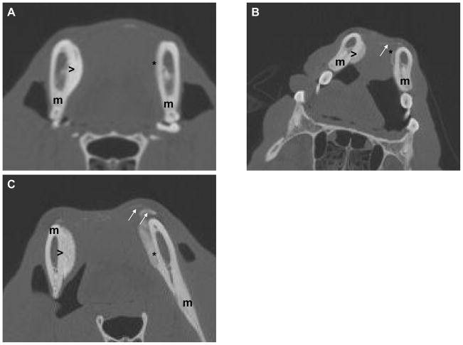
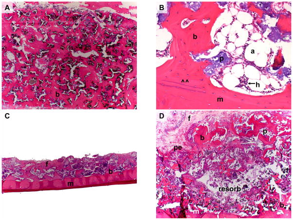
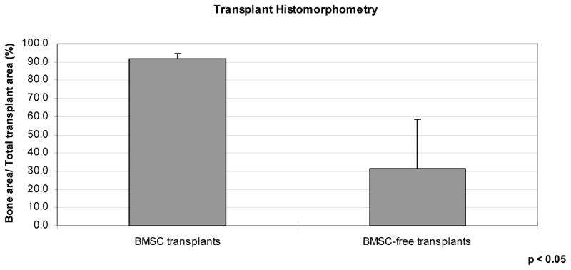
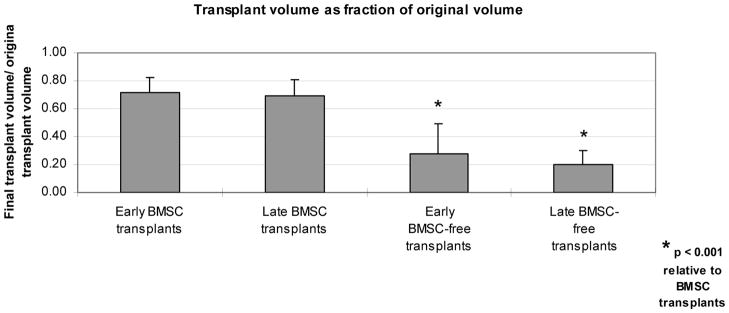
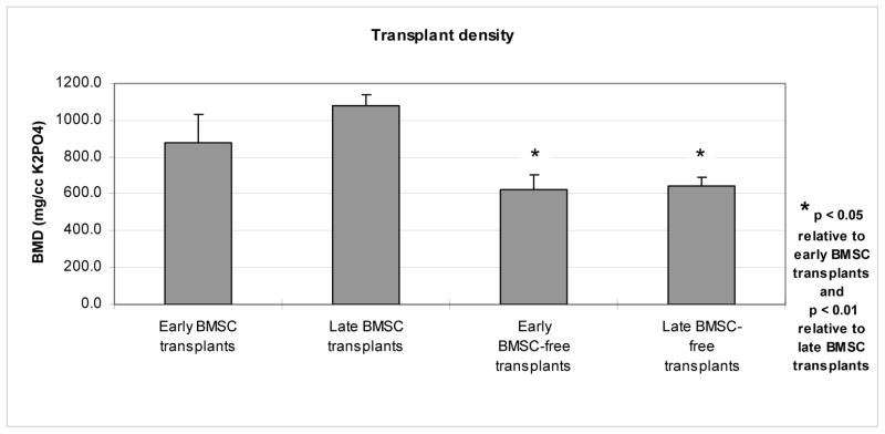
Transplants of culture-expanded bone marrow stromal cells (BMSCs) combined with hydroxyapatite/tricalcium phosphate (HA/TCP) scaffolds successfully form cortico-cancellous bone to reconstruct the dog craniofacial skeleton. Yet, these transplants' long-term stability in large animal models has not been evaluated. This study's purpose was the evaluation of long-term BMSC transplant stability when used to augment the mandible. Here, autologous BMSC-HA/TCP transplants were introduced onto the unilateral dog mandible as onlay grafts, while contralateral control mandibles received HA/TCP onlays alone. Quantitative CT (qCT) scans were obtained both early and late after transplantation. Transplants were harvested up to 19 months later for histologic and mechanical analyses. In all dogs, BMSC transplants formed significantly greater amounts of bone over their control counterparts. The new bone formed an extensive union with the underlying mandible. BMSC transplants retained the majority of their initial volume, while control (HA/TCP only) transplants were nearly completely resorbed. By qCT, the extent of newly formed bone could be determined non-invasively. In summary, HA/TCP particles alone undergo a high degree of resorption, while autologous cultured BMSC-HA/TCP transplants provide long-term bony augmentation of the mandible.


Figures

Figure 1
Figure 1
Figure 1A: CT image of dog #1 at 17 months following transplantation. BMSC transplant (>) exhibits substantial volume and density, and is tightly adherent to underlying mandible (m). BMSC-free transplant (*), in contrast, has undergone nearly complete resorption. Figure 1B: CT image of dog #2 at 15 months following transplantation. Loose HA/TCP particles remain at site of BMSC-free transplant site (arrows). The BMSC transplant retains its volume and density (>). Figure 1C: CT image of dog #3 at 3 months following transplantation. BMSC-free transplant (*) already demonstrates substantial resorption. Loose HA/TCP particles (arrows) have already begun migrating away from the BMSC-free transplant (*). The BMSC transplant (>) exhibits greater density and volume than BMSC-free transplant.
Figure 2
Figure 2
Figure 2A: BMSC-containing transplant (Animal 2) 17 months post-operatively. Note extensive cortico-cancellous bone (b) formed throughout the entire thickness of the transplant. Figure 2B: BMSC-containing transplant (Animal 3) 19 months post-operatively. Note extensive cortico-cancellous bone encircling hematopoiesis (h) and adipocytes (a). Newly formed bone has formed a union (at ^^) with the underlying mandible (m). Figure 2C: BMSC-free transplant (Animal 2) 17 months post-operatively. Note that residual transplant has minimal volume and that much of the original HA/TCP scaffold has been resorbed. Minimal bone (b) and extensive fibrous tissue (f) lay on the mandible (m). Figure 2D: BMSC-free transplant (Animal 3) 19 months post-operatively. Note resorption of transplant interior leaving behind minimal tissue (resorb), and minimal residual bone (b) along mandible (m) and periosteum (pe). (b = bone, f = fibrous connective tissue, p = particle, m = mandible, pe = periosteum, a = adipocyte, h = hematopoiesis), resorb = (resorbed tissue within BMSC-free transplant) Magnification: 2.5x (A, C, D). Magnification: 20x (B). Stain: Hematoxylin and eosin; paraffin embedding following demineralization (A–D)
Figure 3
Figure 3
Extent of bone on histologic tissue sections, as a function of transplant type, assessed via histomorphometry, as a percentage of total transplant cross-sectional area.
Figure 4
Figure 4
Transplant volume on CT examinations, as a function of transplant type and time of imaging, as a percentage of transplant volume at time of transplantation.
Figure 5
Figure 5
Bone mineral density (BMD) on CT examinations, as a function of transplant type and time of imaging.

References

    1. Dado DV, Izquierdo R. Absorption of onlay bone grafts in immature rabbits: membranous versus enchondral bone and bone struts versus paste. Ann Plast Surg. 1989;23:39–48. - PubMed
    1. Mankani MH, Kuznetsov SA, Wolfe RM, Marshall GW, Robey PG. In vivo bone formation by human bone marrow stromal cells: reconstruction of the mouse calvarium and mandible. Stem Cells. 2006;24:2140–9. Epub 2006 Jun 8. - PubMed
    1. Mankani MH, Kuznetsov SA, Shannon B, Nalla RK, Ritchie RO, Qin Y, Robey PG. Canine cranial reconstruction using autologous bone marrow stromal cells. Am J Pathol. 2006;168:542–50. - PMC - PubMed
    1. Krebsbach PH, Mankani MH, Satomura K, Kuznetsov SA, Robey PG. Repair of craniotomy defects using bone marrow stromal cells. Transplantation. 1998;66:1272–8. - PubMed
    1. Kuznetsov SA, Krebsbach PH, Satomura K, Kerr J, Riminucci M, Benayahu D, Robey PG. Single-colony derived strains of human marrow stromal fibroblasts form bone after transplantation in vivo. J Bone Miner Res. 1997;12:1335–47. - PubMed

Publication types