. 2008 Aug 19;105(33):12016-21.
doi: 10.1073/pnas.0802261105. Epub 2008 Aug 7.

Deficiency in ubiquitin ligase TRIM2 causes accumulation of neurofilament light chain and neurodegeneration

Affiliations

Deficiency in ubiquitin ligase TRIM2 causes accumulation of neurofilament light chain and neurodegeneration

Martin Balastik et al. Proc Natl Acad Sci U S A. .

Abstract

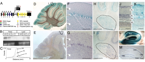
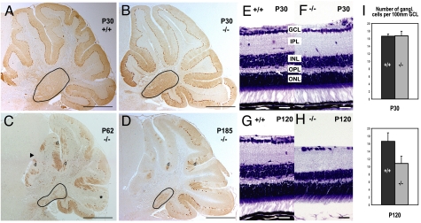
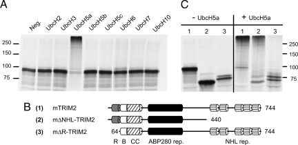
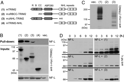
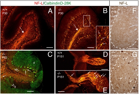
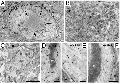
TRIM RING finger proteins have been shown to play an important role in cancerogenesis, in the pathogenesis of some human hereditary disorders, and in the defense against viral infection, but the function of the majority of TRIM proteins remains unknown. Here, we show that TRIM RING finger protein TRIM2, highly expressed in the nervous system, is an UbcH5a-dependent ubiquitin ligase. We further demonstrate that TRIM2 binds to neurofilament light subunit (NF-L) and regulates NF-L ubiquitination. Additionally, we show that mice deficient in TRIM2 have increased NF-L level in axons and NF-L-filled axonal swellings in cerebellum, retina, spinal cord, and cerebral cortex. The axonopathy is followed by progressive neurodegeneration accompanied by juvenile-onset tremor and ataxia. Our results demonstrate that TRIM2 is an ubiquitin ligase and point to a mechanism regulating NF-L metabolism through an ubiquitination pathway that, if deregulated, triggers neurodegeneration.


Conflict of interest statement

The authors declare no conflict of interest.

Figures

Fig. 1.
Fig. 1.
Generation and characterization of TRIM2GT mice. (A) Integration site of the GT vector in the Trim2GT mouse line within the Trim2 locus, inside intron 6. (B) Expression of Trim2 in WT (+/+), Trim2GT heterozygous (+/−), and homozygous (−/−) mice analyzed by Northern blotting with Trim2 3′ UTR probe (Trim2) and a GT vector-specific probe (LacZ) (loading control, ethydium bromide-stained gel). (C) Quantification of Trim2 expression from B by instant imager. (D–M) Trim2 expression in cerebellar Purkinje cells (D–G), deep cerebellar nuclei (H and I, encircled), retina (J and K), and hippocampus (L and M), by β-gal staining of Trim2GT heterozygous mice (D, F, H, K, L) and by in situ hybridization using Trim2 probe (E, G, I, J, M). M, molecular layer; PCL, Purkinje cell layer; GL, granule cell layer; GCL, ganglionic cell layer; IPL, inner plexiform layer; INL, inner nuclear layer; OPL, outer plexiform layer; ONL, outer nuclear layer; Rad, stratum radiatum; Mol, stratum moleculare; DG, dentate gyrus; CA1–3 hippocampal areas (Scale bars: 200 μm in D and E; 100 μm in F–I, L, and M; and 30 μm in J and K.)
Fig. 2.
Fig. 2.
Degeneration in Trim2GT homozygous mice. (A–D) Calbindin D-28K immunostaining of WT (A) and homozygous (B) cerebellar Purkinje cells at postnatal day P30. Degeneration of vermal Purkinje cells and the deep nuclei (encircled) detected in 2- (C) and 6-month-old (D) mutants (anterior lobes, arrowhead; posterior lobe, asterisk; Purkinje cell dendrites, d). (E–H) Nissl staining of retina in WT and Trim2GT homozygous mice at P30 (E and F) and P120 (G and H). Inner nuclear layer, INL; outer plexiform layer, OPL. (I) Quantification of the number of ganglionic cells per 100 μm of the ganglionic cell layer (GCL) in 1- and 4-month-old mice (n = 3). (Scale bars: 700 μm in A–D, 30 μm in E–H.)
Fig. 3.
Fig. 3.
Ubiquitin ligase activity of TRIM2. (A) [35S]Met-labeled TRIM2 autoubiquitination in an in vitro ubiquitination assay in the presence of various ubiquitin-conjugating enzymes (E2). (B) Schematic representation of TRIM2 full-length and mutant constructs. (R, RING finger; B, B box; CC, coiled-coil). (C) In vitro autoubiquitination of full-length and mutant [35S]Met TRIM2 in the absence (−) or presence (+) of UbcH5a.
Fig. 4.
Fig. 4.
TRIM2–NF-L interaction and ubiquitination. (A) Schematic representation of TRIM2 full-length and mutant constructs. (B) NF-L pull-down assay with the distinct TRIM2 constructs in HeLa cells. (C) Autoubiquitination of TRIM2 and its truncated forms in HeLa cells. (D) NF-L ubiquitination time course in HeLa cells—alone (NF-L) and after coexpression of the full-length TRIM2 (NF-L + 1), the ligase-dead mutant (NF-L + 2), or the C-terminally truncated TRIM2 (NF-L + 3).
Fig. 5.
Fig. 5.
Increased NF-L axonal density and NF-L axonal swellings in Trim2GT homozygous mice. (A) WT littermate of B. (B) Trim2GT homozygotes show increased NF-L level at P30 in cerebellar white matter (arrow) and pathological accumulation of NF-L (arrows in the Inset). (C) NF-L-filled axonal swellings colocalize with calbindin D-28K staining of Purkinje cell axons (arrows); d, remaining Purkinje cell dendrites. (E–G) NF-L-filled axonal spheroids (arrows) are also present in the optic nerve (E) and the gray matter of spinal cord (G) of the Trim2GT homozygotes, but not in their WT or heterozygous (D and F) littermates. (Scale bars: 100 μm.)
Fig. 6.
Fig. 6.
Ultrastructure and NF-L immunoelectronmicroscopy of dilated axons in the deep cerebellar nuclei of Trim2GT mice. (A–D) Swollen axons dilated by the accumulation of neurofilaments, microtubuli, mitochondria, and vesicles. (A) Dilated axon with clear signs of degeneration, with the accumulation of dense and multivesicular bodies (arrows) tightly packed (B). In A, numerous regular-size axons can be observed nearby. (C–F) Preembedding immunoelectronmicroscopy using NF-L antibody. Immunometal particles are associated to the disorganized neurofilaments in the dilated axons (C and D) or to the properly oriented parallel neurofilaments in normal axons (E and F). m, mitochondria; my, myelin; dashed line, separation between the axoplasm and the ensheathing myelin. (Scale bars: 2.5 μm in A; μm in C; 500 nm in B; 250 nm in D and E; and 200 nm in F.)

References

    1. Freemont PS. RING for destruction? Curr Biol. 2000;10:R84–R87. - PubMed
    1. Reymond A, et al. The tripartite motif family identifies cell compartments. EMBO J. 2001;20:2140–2151. - PMC - PubMed
    1. de The H, et al. The PML-RAR alpha fusion mRNA generated by the t(15;17) translocation in acute promyelocytic leukemia encodes a functionally altered RAR. Cell. 1991;66:675–684. - PubMed
    1. Kaiser SM, Malik HS, Emerman M. Restriction of an extinct retrovirus by the human TRIM5alpha antiviral protein. Science. 2007;316:1756–1758. - PubMed
    1. Shimura H, et al. Familial Parkinson disease gene product, parkin, is a ubiquitin-protein ligase. Nat Genet. 2000;25:302–305. - PubMed

Publication types