. 2008 Aug 19;105(33):11987-92.
doi: 10.1073/pnas.0804350105. Epub 2008 Aug 7.

Spinocerebellar ataxia type 6 knockin mice develop a progressive neuronal dysfunction with age-dependent accumulation of mutant CaV2.1 channels

Affiliations

Spinocerebellar ataxia type 6 knockin mice develop a progressive neuronal dysfunction with age-dependent accumulation of mutant CaV2.1 channels

Kei Watase et al. Proc Natl Acad Sci U S A. .

Abstract

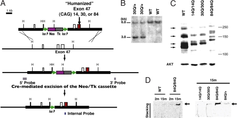
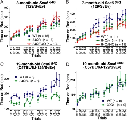
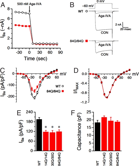
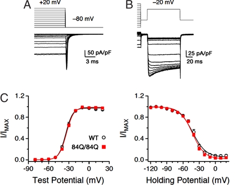
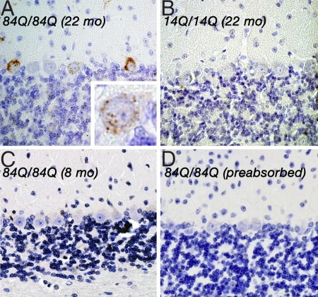
Spinocerebellar ataxia type 6 (SCA6) is a neurodegenerative disorder caused by CAG repeat expansions within the voltage-gated calcium (Ca(V)) 2.1 channel gene. It remains controversial whether the mutation exerts neurotoxicity by changing the function of Ca(V)2.1 channel or through a gain-of-function mechanism associated with accumulation of the expanded polyglutamine protein. We generated three strains of knockin (KI) mice carrying normal, expanded, or hyperexpanded CAG repeat tracts in the Cacna1a locus. The mice expressing hyperexpanded polyglutamine (Sca6(84Q)) developed progressive motor impairment and aggregation of mutant Ca(V)2.1 channels. Electrophysiological analysis of cerebellar Purkinje cells revealed similar Ca(2+) channel current density among the three KI models. Neither voltage sensitivity of activation nor inactivation was altered in the Sca6(84Q) neurons, suggesting that expanded CAG repeat per se does not affect the intrinsic electrophysiological properties of the channels. The pathogenesis of SCA6 is apparently linked to an age-dependent process accompanied by accumulation of mutant Ca(V)2.1 channels.


Conflict of interest statement

The authors declare no conflict of interest.

Figures

Fig. 1.
Fig. 1.
Generation of Sca6 KI mice. (A) Targeting scheme represents the targeting construct, the endogenous Sca6 allele, and the predicted structure of the mutant Sca6 allele generated by a homologous recombination and a Cre-mediated excision event. (B) Germ-line transmission of a Sca630Q allele. Southern blot analysis of HindIII-digested tail DNA revealed 6.8-kb WT and 3.8-kb mutant bands in Sca630Q/30Q mice with the internal probe shown in A. (C) Western blot analysis performed on cytoplasmic fraction of cerebellar lysates from 6-month-old Sca614Q/14Q, Sca630Q/30Q, and Sca684Q/84Q KI mice and a 6-month-old WT mouse, all blotted against CT-2 (Upper) and AKT (Lower, used as a loading control). Arrows indicate multiple CT-2 positive IRs detected with WT cerebellar lysates. Mutant CaV2.1 shows less mobility because of the polyglutamine stretch and its flanking human-specific sequence. (D) Detection of aggregated CaV2.1 channel in Sca684Q cerebella. Shown are immunoblots of cerebellar extracts from 2- (2m) or 15-month-old (15m) Sca614Q/14Q (14Q/14Q), Sca6300Q/30Q, Sca684Q/+, Sca6840Q/84Q KI, and WT mice. Arrow indicates CT-2 IRs detected in the stacking part of the gels. Molecular masses are indicated at the right of each panel in kDa.
Fig. 2.
Fig. 2.
Analysis of the Sca6 KI mice on the accelerating Rotarod apparatus. (A and B) Performance of 3- (A) and 7-month-old (B) Sca684Q84Q mice of 129/SvEv background. Sca684Q/84Q showed impaired motor performance (P = 0.035) at 7 months of age. (C) Performance of 19-month-old heterozygous Sca684Q/+ mice of C57BL/6J-129/SvEv background. (D) Performance of 19-month-old heterozygous Sca630Q/+ mice of C57BL/6J-129/SvEv background. Mice were trained in four trials per day (T1–T4) for four days (D1–D4). Error bars indicate SEM.
Fig. 3.
Fig. 3.
Whole-cell Purkinje Ba2+ currents are blocked by Aga-IVA and reduced in Sca6 neurons. (A) Time course of peak current for a WT (○) or an Sca684Q84Q (■) neuron. Aga-IVA was added to the bath when indicated, to a final bath concentration of ≈500 nM. (B) Individual traces from the two cells in A, before and after bath application of Aga-IVA. (C–F) Each group consists of 4–19 neurons. (C) Current density was plotted against voltage for WT (○) and Sca6 84Q/84Q (■) neurons. (D) Same data as in A, normalized to peak inward current. (E) Summary of whole-cell current density at −15 mV. *, P < 0.005 vs. WT (by two-tailed Student's unpaired t test). (F) Summary of whole-cell capacitance. P > 0.05 vs. WT for all three Sca6 populations. 14Q/14Q, 30Q/30Q, and 84Q/84Q represent Sca614Q/14Q, Sca630Q/30Q, and Sca684Q/84Q, respectively.
Fig. 4.
Fig. 4.
Sca6 mutations do not affect activation or inactivation. (A) Protocol used to measure activation (G–V) curves. Shown are currents from a WT PC. The cell was held at −80 mV and depolarized for 60 ms to incremental voltages from −80 through +20 mV. Peak tail current amplitude was normalized and plotted against voltage to generate the curves in C. (Scale bars: vertical, 50 pA/pF; horizontal, 3 ms.) (B) Protocol used to measure steady-state inactivation for the same cell shown in A. The cell was held at −80 mV, and a 3-s conditioning pulse was applied in 10-mV increments from −120 through +20 mV followed by a test pulse to −20 mV. Peak current amplitude during the test pulse was normalized and plotted against conditioning pulse voltage to generate the curves in C. (C) Summary of activation and inactivation curves for WT (○) and Sca684Q84Q (■) PCs. Solid lines are Boltzmann fits of the data. For activation, WT and Sca684Q/84Q V0.5 are −34.0 ± 0.9 and −33.1 ± 0.5 mV, respectively (P >0.4, by two-tailed Student's unpaired t test); slope factors (k) are 4.8 ± 0.2 and 4.8 ± 0.1, respectively (P > 0.9). For inactivation, WT and Sca684Q/84Q V0.5 are −41.8 ± 1.0 and −41.6 ± 0.8 mV, respectively (P > 0.8); slope factors (k) are 10.0 ± 0.7 and 9.3 ± 0.5, respectively (P > 0.4). n = 10–13 neurons.
Fig. 5.
Fig. 5.
NI formation in Sca684Q84Q Purkinje neurons. (A) Immunohistochemistry for A6RPT-polyQ revealed NI formation in Sca684Q/84Q PCs (22-month-old). (Inset) Individual PC at higher magnification. Note that these NIs were seen mainly in the cytoplasm of these neurons. Similar NI-harboring PCs were scattered in the cerebellum. (B and C) In contrast, only weak immunoreactivities were detected in the cerebellum of a 22-month-old Sca614Q/14Q (B) or an 8-month-old Sca684Q/84Q mouse (C). (D) The immunoreactivity was entirely blocked in the cerebellum of the 22-month-old Sca684Q/84Q mouse when A6RPT-polyQ was preincubated with its polypeptide antigens. (Magnification of original photographs, ×400.) Parallel staining undertaken with control human specimen also revealed similar NI formation in SCA6 PCs (data not shown).

References

    1. Zhuchenko O, et al. Autosomal dominant cerebellar ataxia (SCA6) associated with small polyglutamine expansions in the α1A-voltage dependent calcium channel. Nat Genet. 1997;15:62–69. - PubMed
    1. Ishikawa K, et al. Japanese families with autosomal dominant pure cerebellar ataxia map to chromosome 19p13.1-p13.2 and are strongly associated with mild CAG expansions in the spinocerebellar ataxia type 6 gene in chromosome19p13.1. Am J Hum Genet. 1997;61:336–346. - PMC - PubMed
    1. Tsuchiya K, et al. Degeneration of the inferior olive in spinocerebellar ataxia 6 may depend on disease duration: Report of two autopsy cases and statistical analysis of autopsy cases reported to date. Neuropathology. 2005;25:125–135. - PubMed
    1. Gatchel JR, Zoghbi HY. Diseases of unstable repeat expansion: Mechanisms and common principles. Nat Rev Genet. 2005;6:743–755. - PubMed
    1. Mariotti C, et al. Pathogenic effect of an intermediate-size SCA6-allele (CAG)(19) in a homozygous patient. Neurology. 2001;57:1502–1504. - PubMed

Publication types

MeSH terms

Substances