. 2008 Nov 15;112(10):4158-69.
doi: 10.1182/blood-2008-02-140814. Epub 2008 Aug 7.

Wiskott-Aldrich syndrome protein deficiency in B cells results in impaired peripheral homeostasis

Affiliations

Wiskott-Aldrich syndrome protein deficiency in B cells results in impaired peripheral homeostasis

Almut Meyer-Bahlburg et al. Blood. .

Abstract

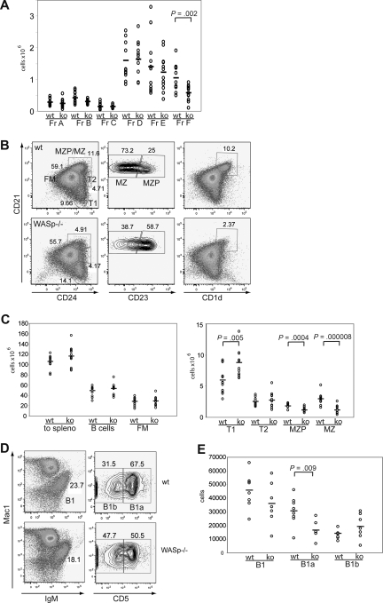
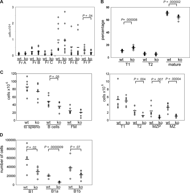
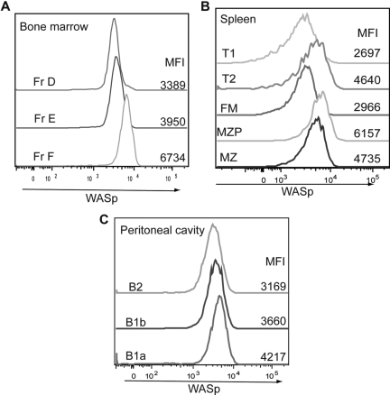
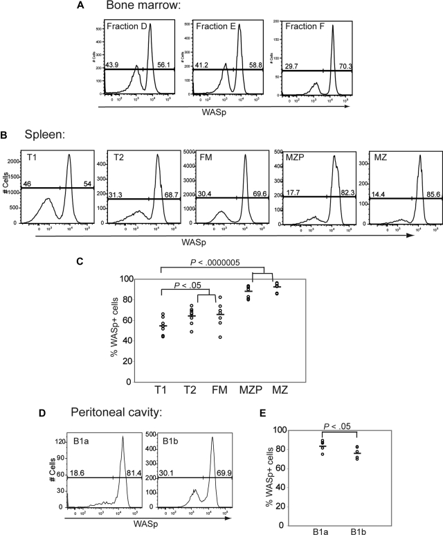
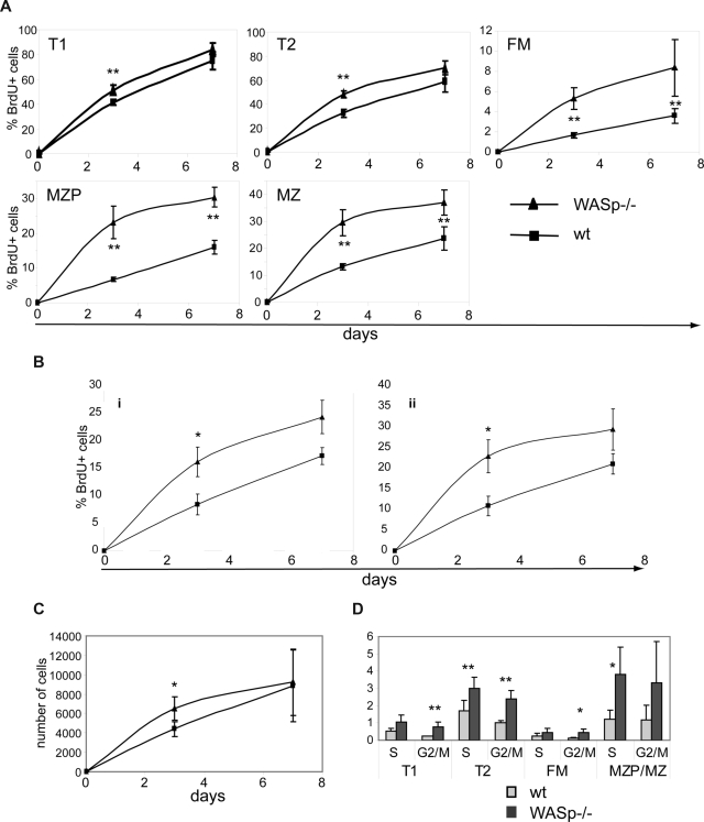
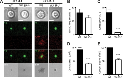
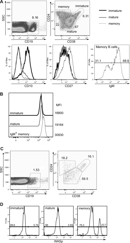
To more precisely identify the B-cell phenotype in Wiskott-Aldrich syndrome (WAS), we used 3 distinct murine in vivo models to define the cell intrinsic requirements for WAS protein (WASp) in central versus peripheral B-cell development. Whereas WASp is dispensable for early bone marrow B-cell development, WASp deficiency results in a marked reduction in each of the major mature peripheral B-cell subsets, exerting the greatest impact on marginal zone and B1a B cells. Using in vivo bromodeoxyuridine labeling and in vitro functional assays, we show that these deficits reflect altered peripheral homeostasis, partially resulting from an impairment in integrin function, rather than a developmental defect. Consistent with these observations, we also show that: (1) WASp expression levels increase with cell maturity, peaking in those subsets exhibiting the greatest sensitivity to WASp deficiency; (2) WASp(+) murine B cells exhibit a marked selective advantage beginning at the late transitional B-cell stage; and (3) a similar in vivo selective advantage is manifest by mature WASp(+) human B cells. Together, our data provide a better understanding of the clinical phenotype of WAS and suggest that gene therapy might be a useful approach to rescue altered B-cell homeostasis in this disease.


Figures

Figure 1
Figure 1
The B-cell phenotype in WASp-deficient mice. (A) BM was stained for expression of B220, CD43, IgM, BP1, CD24, CD19, and IgD and developmental stages determined according to the Hardy classification. Absolute numbers for each fraction (Fr) A through F are shown in WT versus WASp−/− mice (ko) (n = 12). (B,C) Splenocytes were stained for expression of B220, CD19, CD24, CD21, CD23, and CD1d and B-cell subsets analyzed. (B) Representative FACS blot gated on B220+CD19+ cells in a WT versus WASp−/− mouse. (C) Absolute numbers of splenic B-cell subsets from WT versus WASp−/− mice (n = 12). (D,E) Peritoneal B cells were stained for IgM, CD5, and Mac1. (D) Representative FACS blot of gating B1 cells in a WT and a WASp−/− mouse. (E) Absolute numbers of B1, B1a, and B1b cells in WT (n = 8) versus WASp−/− mice (n = 7).
Figure 2
Figure 2
The WASp-deficient B-cell phenotype is B cell–intrinsic. BM from WT or WASp−/− mice was transplanted into sublethally irradiated μMT mice. Recipient mice were killed 6 weeks after transplantation, and B-cell subsets in BM, spleen, and peritoneal cavity were analyzed. Shown are absolute numbers for (A) BM (n = 9 recipient mice), (C) spleen (n = 7), and (D) peritoneal cavity (n = 7) and the relative percentage of subsets in (B) peripheral blood (n = 12).
Figure 3
Figure 3
Relative WASp expression levels in distinct B-cell subsets. B cells in different lymphoid compartments from a WT mouse were stained for WASp expression in addition to surface markers to determine B-cell subpopulations. Relative WASp expression and mean fluorescent intensity are shown for each of the B-cell subsets derived from (A) BM, (B) spleen, and (C) peritoneal cavity. Data shown are representative of more than 5 individual mice.
Figure 4
Figure 4
WASp+ B cells exhibit a selective advantage beginning at the late transitional B-cell stage. Expression of WASp was determined in B-cell subsets derived from BM, spleen, and peritoneal cavity in female WASp heterozygous mice. Representative example of staining in (A) BM, (B) spleen, and (D) peritoneal cavity. Percentage of WASp-positive B cells in each subset in (C) spleen (n = 8) and (E) peritoneal cavity (n = 6).
Figure 5
Figure 5
B cells in WASp-deficient mice exhibit a higher turnover rate. (A-C) WT and WASp−/− mice were continuously fed BrdU via the drinking water for up to 7 days. Mice were killed on day 3 or 7 and splenocytes stained for BrdU in addition to surface makers to identify B-cell subsets. Percentage of BrdU+ cells in (A) splenic and (B) peritoneal B-cell subsets. (C) Absolute numbers of BrdU+ MZ B cells in WT versus WASp−/− mice. Data shown reflect the average of 3 mice per time point and are representative of 2 independent experiments. *P ≤ .05. (D) For cell-cycle analysis, splenocytes from WT or WASp−/− mice were stained with PyroninY and DAPI in addition to surface markers. Data show the percentage of cells in either S- or G2/M phase of the cell cycle (average of 3 mice) from one of 3 independent experiments. *P ≤ .05; **P ≤ .01.
Figure 6
Figure 6
WASp is required for integrin activation and pSMAC formation at the B-cell immunologic synapse. WT and WASp−/− B cells were settled onto lipid bilayers containing anti-kappa antibody (green) or antikappa and Alexa532-conjugated ICAM-1 (red) and imaged by confocal microscopy after 20 minutes of interaction. (A) Differential interference contrast (DIC), fluorescence and interference reflection microscopy (IRM) images of representative cells are shown. Quantification of the percentage of B cells forming (B) a cSMAC or (C) an IS (cSMAC + pSMAC) and (D,E) the area of B-cell contact with the bilayer. For each condition, a total of 20 to 50 B cells were analyzed. Bars represent mean values plus or minus SEM. Data are representative of 2 independent experiments. Scale bar represents 2.5 μm. ***P < .001.
Figure 7
Figure 7
WASp+ mature B cells exhibit a selective advantage in humans. (A,B) Expression of WASp in immature, mature, and IgM memory B cells in peripheral blood from a healthy person. Peripheral blood mononuclear cells from a healthy person were stained for CD19, CD24, CD38, CD10, CD27, and IgM in addition to WASp. (A) Based on these markers, the following B-cell subsets were identified: CD19+CD24hiCD38hi immature/transitional B cells, CD19+CD24lowCD38int naive mature B cells, and CD19+CD24hiCD38lowIgM+ IgM memory B cells. Transitional B cells express high levels of CD10, whereas memory B cells express high levels of CD27. (B) WASp expression and relative mean fluorescent intensity are shown for each of these B-cell subsets. Data shown are representative of more than 3 healthy persons. (C,D) Analysis of B cells in a WAS patient with a revertant mutation. B-cell subsets and relative WASp expression were determined in a WAS patient with a known revertant mutation. (C) B-cell subsets were identified as described for panel A. (D) WASp expression in immature, mature, and memory B cells (gated as in panel A) from the WAS patient.

Comment in

  • WASp stings mature lymphocytes.
    Severinson E. Severinson E. Blood. 2008 Nov 15;112(10):3921-2. doi: 10.1182/blood-2008-09-176685. Blood. 2008. PMID: 18988873 Free PMC article.

References

    1. Sullivan KE, Mullen CA, Blaese RM, Winkelstein JA. A multiinstitutional survey of the Wiskott-Aldrich syndrome. J Pediatr. 1994;125:876–885. - PubMed
    1. Ochs HD, Slichter SJ, Harker LA, Von Behrens WE, Clark RA, Wedgwood RJ. The Wiskott-Aldrich syndrome: studies of lymphocytes, granulocytes, and platelets. Blood. 1980;55:243–252. - PubMed
    1. Park JY, Kob M, Prodeus AP, Rosen FS, Shcherbina A, Remold-O'Donnell E. Early deficit of lymphocytes in Wiskott-Aldrich syndrome: possible role of WASP in human lymphocyte maturation. Clin Exp Immunol. 2004;136:104–110. - PMC - PubMed
    1. Ochs HD, Thrasher AJ. The Wiskott-Aldrich syndrome. J Allergy Clin Immunol. 2006;117:725–738. quiz 739. - PubMed
    1. Thrasher AJ. WASp in immune-system organization and function. Nat Rev Immunol. 2002;2:635–646. - PubMed

Publication types

Substances