. 2008 Sep;173(3):700-15.
doi: 10.2353/ajpath.2008.071128. Epub 2008 Aug 7.

Endometrial-peritoneal interactions during endometriotic lesion establishment

Affiliations

Endometrial-peritoneal interactions during endometriotic lesion establishment

M Louise Hull et al. Am J Pathol. 2008 Sep.

Abstract

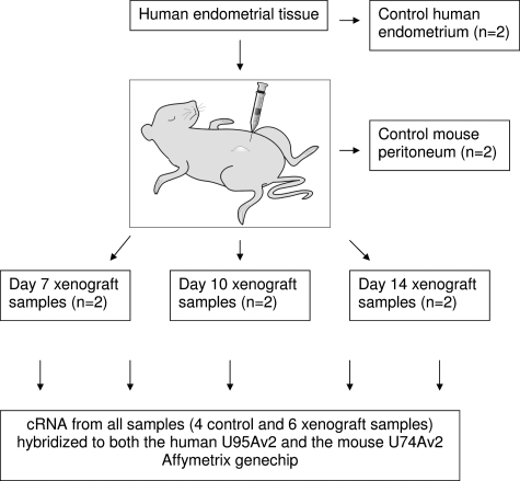
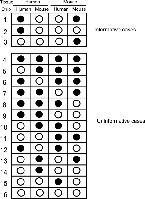
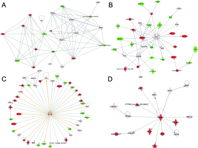
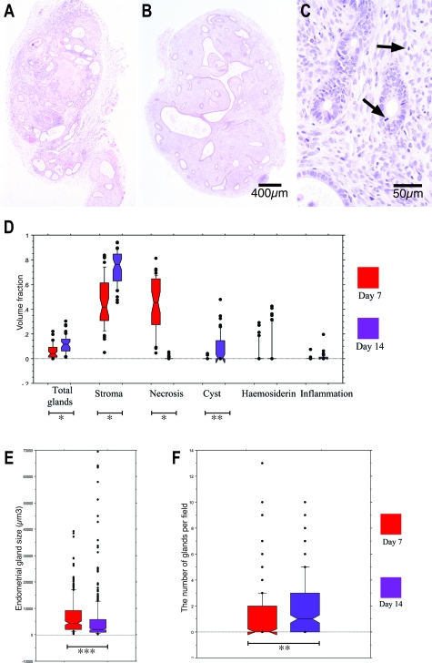
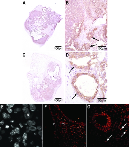
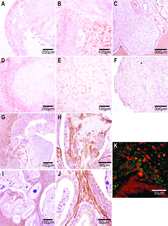
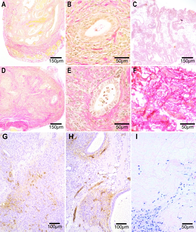
The pathophysiology of endometriosis remains unclear but involves a complex interaction between ectopic endometrium and host peritoneal tissues. We hypothesized that disruption of this interaction would suppress endometriotic lesion formation. We hoped to delineate the molecular and cellular dialogue between ectopic human endometrium and peritoneal tissues in nude mice as a first step toward testing this hypothesis. Human endometrium was xenografted into nude mice, and the resulting lesions were analyzed using microarrays. A novel technique was developed that unambiguously determined whether RNA transcripts identified via microarray analyses originated from human cells (endometrium) or mouse cells (mesothelium). Four key pathways (ubiquitin/proteasome, inflammation, tissue remodeling/repair, and ras-mediated oncogenesis) were revealed, demonstrating communication between host mesothelial cells and ectopic endometrium. Morphometric analysis of nude mouse lesions confirmed that necrosis, inflammation, healing and repair, and cell proliferation occurred during xenograft development. These processes were entirely consistent with the molecular networks revealed by the microarray data. The transcripts detected in the xenografts overlapped with differentially expressed transcripts in a comparison between paired eutopic and ectopic endometria from human endometriotic patients. For the first time, components of the interaction between ectopic endometrium and peritoneal stromal tissues are revealed. Targeted disruption of this dialogue is likely to inhibit endometriotic tissue formation and may prove to be an effective therapeutic strategy for endometriosis.


Figures

Figure 1
Figure 1
Diagram illustrating the source of tissue for all control and xenograft samples. Each xenograft sample was pooled from three to five nude mouse lesions at each time point. cDNA from each sample was hybridized to both the HU-95A (human) and MU-74-Av2 (murine) Affymetrix GeneChip.
Figure 2
Figure 2
Schematic representation of the possible outcomes of hybridizing human cRNA and mouse cRNA to both human and mouse arrays. Each probe set on the array will produce one of the 16 possible hybridization patterns. Filled circles represent positive hybridization. Some probe sets hybridize with cRNAs derived from both species (ie, cross-hybridize, for example cases 9 and 11); others are uninformative as there is no signal from the hybridization with the correct species (for example cases 14–16).
Figure 3
Figure 3
Four key Ingenuity Pathways Analyses (A–D) representing the molecular relationships between the unambiguously identified human and mouse transcripts in nude mouse xenografts. Each node represents a transcript: green, mouse-specific; red, human-specific; pink, represented in both human and mouse transcriptomes. Supplemental Table 3 at http://ajp.amjpathol.org lists the gene names for all abbreviations. The gene products are represented as nodes, and the biological relationship between two nodes is represented as a line. All lines are supported by at least one reference in literature, textbook, or from canonical information stored in the Ingenuity Pathways knowledge base.
Figure 4
Figure 4
A–C: Formalin-fixed sections stained with hematoxylin and eosin from day 7 (A) and day 14 (B and C) nude mouse xenografts. A: Day 7 nude mouse xenografts exhibited central necrosis, glandular degeneration, and leukocyte infiltration. B: Day 14 implants contained an epithelialized centralized cystic space surrounded by multiple pseudostratified glands. C: Mitotic figures are identified in day 14 glandular and stromal cells (arrows). D: Volume fraction analysis of day 7 and day 14 nude mouse explants. Necrosis occupied a larger volume of day 7 lesions, whereas a higher volume fraction of total glands, stroma, and cysts was present in day 14 xenografts (*P < 0.0001; **P < 0.001). E: Two-dimensional nucleation endometrial gland size estimates from day 7 and day 14 nude mouse lesions. Endometrial gland size was significantly reduced in day 14 lesions (*P < 0.0001). F: The number of glands per field in day 7 and day 14 nude mouse xenografts. There were more glands per field in day 14 xenografts (***P = 0.002).
Figure 5
Figure 5
A–D: Immunohistochemical localization of human MHC class I antigens in frozen sections from day 7 (A and B) and day 14 (C and D) nude mouse lesions. A and B: In day 7 lesions, centrally located glandular epithelium and stroma demonstrate intense brown human MHC class I immunoreactivity whereas unstained murine cells are seen around the circumference of the lesion (A) and in tongues extending into the center (B). C and D: In day 14 xenografts, anti-human MHC class I antibody staining is more diffuse (C) with significant numbers of unstained murine cells seen in central locations of the xenografts (D). The glandular epithelium maintains strong human MHC class I immunoreactivity in day 7 (B, arrows) and day 14 (D, arrows) lesions. E–G: Fluorescent staining of formalin-fixed nude mouse xenografts using the Hoechst reagent. E: A gray scale high-magnification view shows cells with uniformly Hoechst-stained nuclei (h, human) immediately adjacent to cells with speckled nuclei (m, murine). F: In a day 7 lesion, the glow over function demonstrates the punctate nuclei of murine stromal cells (m) between the homogeneously nucleated human cells of two glandular epithelia (h). G: Punctate solitary murine nuclei (arrows) can be seen completely surrounded by cells with homogenous human nuclei in the central part of a day 14 lesion.
Figure 6
Figure 6
Antibody reactivity in frozen sections from day 7 (A–C) and day 14 (D–F) nude mouse xenografts. A and B: Many brown-stained, F4/80-positive murine macrophages are identified in the peripheral rim of frozen sections from day 7 xenografts (A) and between peripheral glandular epithelia (B). D and E: Large numbers of brown F4/80-positive murine macrophages are present in the periphery of day 14 nude mouse lesions (D). F4/80-positive macrophages are less densely scattered throughout the stroma of central portions of the day 14 xenografts (E). C and F: Anti-human CD68 immunoreactivity in paraffin-embedded nude mouse lesions. There were no CD68-positive cells in paraffin-embedded sections from day 7 (C) or day 14 (F) nude mouse xenografts. G–J: Immunohistochemical identification of α-SMA-positive cells in paraffin-embedded sections from day 7 (G and H) and day 14 (I and J) nude mouse xenografts. Large numbers of brown-stained, α-SMA-positive myofibroblasts are present in the outer third of paraffin-embedded sections from day 7 nude mouse lesions (G), and some are immediately adjacent to the necrotic core (H). Spindle-shaped α-SMA-positive myofibroblasts are seen subepithelially in the stromal tissue in central parts of the day 14 xenografts (I and J). K: Fluorescent colocalization of anti-α-SMA antibodies and the Hoechst nuclear stain in a formalin-fixed day 14 nude mouse lesion revealed green α-SMA-positive fibers in the cytoplasm of subepithelial cells that contain orange speckled Hoechst-stained (murine) nuclei. The glandular epithelium is made up of cells with a homogeneous (ie, human) nuclear appearance after Hoechst staining.
Figure 7
Figure 7
A–F: Staining of collagen fibers using Weigert’s iron hematoxylin and Van Gieson’s method in formalin-fixed day 7 (A and B) and day 14 (D and E) nude mouse lesions and in frozen sections of human eutopic (C) and ectopic (F) endometrium from the same volunteer. Sparse red collagen fibers are present at the edges of formalin-fixed day 7 nude mouse lesions (A) and in more central locations by day 14 (D). The collagen fibers encircle the endometrial glands between day 7 (B) and 14 (E). Eutopic human endometrium (C) displays light red collagen staining, whereas ectopic endometrium (F) exhibits intense collagen staining. G and H: Von Willebrand factor (vWF) immunoreactivity in formalin-fixed sections from day 7 (G) and 14 (H) nude mouse lesions. Scattered brown vWF vascular endothelial cells are present in the periphery of day 7 xenografts (G), whereas small vessels made up of brown endothelial cells are present in central locations of day 14 lesions (H). I: Hematoxylin and eosin staining of a day 7 nude mouse lesion show purple-stained cells lined up along fibers of the extracellular matrix.

References

    1. Strathy JH, Molgaard CA, Coulam CB, Melton LJ., 3rd Endometriosis and infertility: a laparoscopic study of endometriosis among fertile and infertile women. Fertil Steril. 1982;38:667–672. - PubMed
    1. Giudice LC, Kao LC. Endometriosis. Lancet. 2004;364:1789–1799. - PubMed
    1. Sampson JA. Peritoneal endometriosis due to menstrual dissemination of endometrial tissue into the peritoneal cavity. Am J Obstet Gynecol. 1927;14:442–469.
    1. Bulun SE, Yang S, Fang Z, Gurates B, Tamura M, Sebastian S. Estrogen production and metabolism in endometriosis. Ann NY Acad Sci. 2002;955:75–85. - PubMed
    1. Ho HN, Wu MY, Yang YS. Peritoneal cellular immunity and endometriosis. Am J Reprod Immunol. 1997;38:400–412. - PubMed

Publication types