. 2008 Sep;173(3):824-34.
doi: 10.2353/ajpath.2008.071005. Epub 2008 Aug 7.

Paracrine overexpression of insulin-like growth factor-1 enhances mammary tumorigenesis in vivo

Affiliations

Paracrine overexpression of insulin-like growth factor-1 enhances mammary tumorigenesis in vivo

Krisztina Kovács de Ostrovich et al. Am J Pathol. 2008 Sep.

Abstract

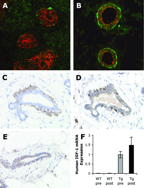
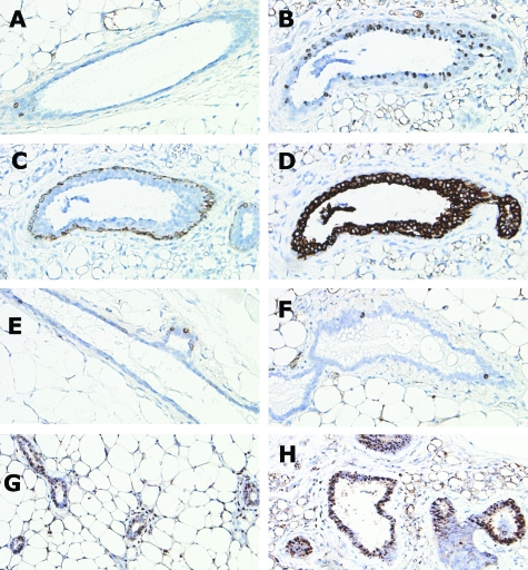
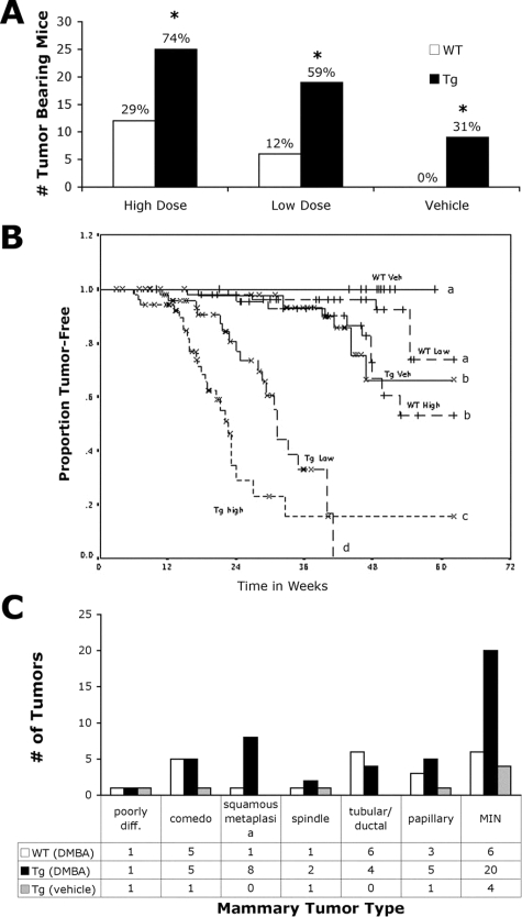
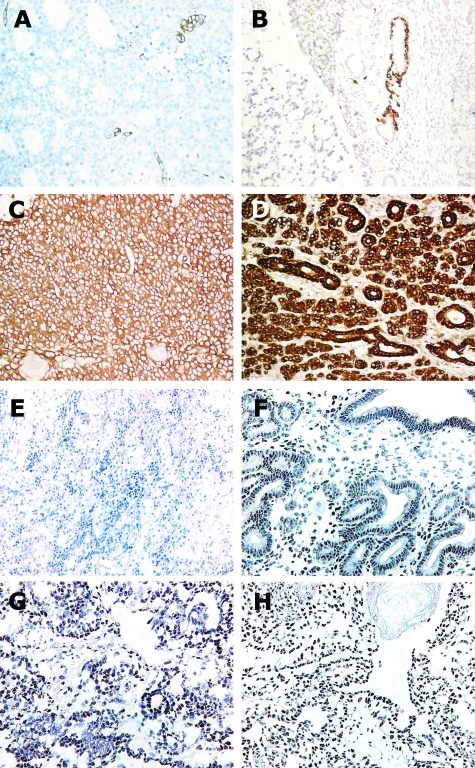
Insulin-like growth factor-1 (IGF-1) stimulates proliferation, regulates tissue development, protects against apoptosis, and promotes the malignant phenotype in the breast and other organs. Some epidemiological studies have linked high circulating levels of IGF-1 with an increased risk of breast cancer. To study the role of IGF-1 in mammary tumorigenesis in vivo, we used transgenic mice in which overexpression of IGF-1 is under the control of the bovine keratin 5 (BK5) promoter and is directed to either the myoepithelial or basal cells in a variety of organs, including the mammary gland. This model closely recapitulates the paracrine exposure of breast epithelium to stromal IGF-1 seen in women. Histologically, mammary glands from transgenic mice were hyperplastic and highly vascularized. Mammary glands from prepubertal transgenic mice had significantly increased ductal proliferation compared with wild-type tissues, although this difference was not maintained after puberty. Transgenic mice also had increased susceptibility to mammary carcinogenesis, and 74% of the BK5.IGF-1 mice treated with 7,12-dimethylbenz[a]anthracene (20 microg/day) developed mammary tumors compared with 29% of the wild-type mice. Interestingly, 31% of the vehicle-treated BK5.IGF-1 animals, but none of the wild-type animals, spontaneously developed mammary cancer. The mammary tumors were moderately differentiated adenocarcinomas that expressed functional, nuclear estrogen receptor at both the protein and mRNA levels. These data support the hypothesis that tissue overexpression of IGF-1 stimulates mammary tumorigenesis.


Figures

Figure 1
Figure 1
K5, K8, and IGF-1 transgene expression in mammary glands. Dual immunofluorescent detection of K5 (green) and K8 (red) antigens in mammary glands from female wild-type perinatal mice, sacrificed on the day of birth, day 0 (A) and at 2.5 days old (B). Serial sections of a postpubertal BK5.IGF-1 transgenic mammary gland immunostained for K5 (C) and IGF-1 (D). Dark staining of IGF-1 was seen in the K5-positive myoepithelial cells. In comparison, there was only light IGF-1 immunostaining in the ductal cells of postpubertal wild-type mammary gland (E). Human IGF-1 mRNA expression was determined in wild-type (WT) and BK5.IGF-1 transgenic (Tg) pre- and postpubertal mammary glands by QPCR (n = 6 for each group) (F). As expected, there was no detectable human IGF-1 mRNA in wild-type glands, but human IGF-1 mRNA was expressed in both pre- and postpubertal transgenic mammary glands. All mRNA levels were normalized to the expression of TATA box binding protein mRNA as described in Materials and Methods. Original magnifications: A and B, ×60; C–E, ×40.
Figure 2
Figure 2
Whole mounts and histological sections from wild-type and BK5.IGF-1 transgenic mice. Whole mounts of mammary gland from 5-week-old wild-type mouse (A) and from 5-week-old BK5.IGF-1 transgenic mouse (B). H&E-stained sections of gland from virgin, adult (postpubertal) wild-type mouse (C) and from age-matched, virgin BK5.IGF-1 transgenic mouse (D). Toluidine blue staining for mast cells in wild-type (E) and BK5.IGF-1 transgenic (F) postpubertal mammary glands. Arrows in F indicate dark-staining mast cells in the transgenic gland. Blood vessels present in H&E-stained sections demonstrate increased vascularity in postpubertal mammary gland of transgenic mice (H) compared to wild-type gland (G). Original magnifications: A and B, ×1; C and D, ×10; E–H, ×40.
Figure 3
Figure 3
Immunohistochemical expression of K5, K8, BrdU, and phospho-Akt in sections of mammary glands from wild-type and BK5.IGF-1 transgenic mice. BrdU immunostaining in wild-type prepubertal gland (A). Serial sections of BK5.IGF-1 transgenic prepubertal gland demonstrating immunolocalization of BrdU (B), K5 (C), and K8 (D). There were more BrdU positive cells in the prepubertal transgenic gland and most of the BrdU positive cells were also K8 positive. BrdU immunostaining of postpubertal wild-type (E) and transgenic (F) glands was not significantly different. Immunostaining for phospho-Akt in prepubertal wild-type (G) and transgenic (H) glands. Phospho-Akt staining was more intense in the ductal epithelial cells of transgenic compared to wild-type glands. Original magnifications, ×40.
Figure 4
Figure 4
Incidence, latency and histological classification of mouse mammary tumors in the BK5.IGF-1 model. Tumor incidence in transgenic (Tg) and wild-type (WT) mice treated with corn oil vehicle or DMBA at 20 μg/mouse/day (high dose) or 10 μg/mouse/day (low dose) (A). Actual numbers of tumor-bearing mice are graphed with the percentage of tumor-bearing mice shown above each bar. The final numbers of mice in each arm were: WT, 41 in high dose, 50 in low dose, and 52 in vehicle only groups; Tg, 34 in high dose, 32 in low dose, and 30 in vehicle only groups. *P < 0.001 versus similarly treated wild-type mice (Fisher’s exact test). Kaplan-Meier analysis of tumor development in all treatment groups (B). Veh, vehicle only. Curves denoted by different letters are significantly different, P < 0.02 (log rank test). Because some mice died or were sacrificed due to other health problems (especially thymoma development in transgenic mice) before termination of the experiment at 55 weeks, final proportions of tumor-free mice do not exactly match the percent incidence values in A. Graphical representation showing the number of each histological tumor type that developed in wild-type mice treated with DMBA [WT (DMBA)], in transgenic mice treated with DMBA [Tg (DMBA)] and in transgenic mice treated with vehicle only [Tg (vehicle)] (C). Poorly diff., poorly differentiated; MIN, mammary intraepithelial neoplasia.
Figure 5
Figure 5
Histological subtypes of mammary tumors. Photomicrographs of H&E-stained sections from DMBA-treated BK5.IGF-1 transgenic and wild-type mice. The ductal phenotype (A) was seen most commonly in tumors from wild-type animals, while transgenic tumors frequently had areas of squamous metaplasia and keratinization (B). Relatively more comedo-type tumors (C) were seen in wild-type rather than transgenic mice. Original magnifications: A and C, ×10; B, ×20.
Figure 6
Figure 6
Immunohistochemical detection of K5 (A, B) and K8 (C, D) antigens in mammary tumors from DMBA-treated wild-type (A, C) and vehicle-treated BK5.IGF-1 transgenic (B, D) mice. Immunostaining of ER in mammary tumors from BK5.IGF-1 transgenic and wild-type mice. Negative (kidney) (E) and positive (uterus) (F) tissue controls. Representative sections of ER immunolocalization in mammary adenocarcinomas from a wild-type (G) and a BK5.IGF-1 transgenic (H) mouse. Original magnifications: A–E, ×10; F–H, ×20.
Figure 7
Figure 7
ER, progesterone receptor, and cyclin D1 expression in tissues and tumors, measured by QPCR (A–C) and Western analysis (D). ER mRNA expression in whole postpubertal mammary glands and mammary tumors from wild-type (WT) and BK5.IGF-1 transgenic (Tg) mice (A). Progesterone receptor mRNA expression in tumors (average of 10 different samples) from untreated wild-type and transgenic mice and in tumors from one wild-type and one transgenic mouse 6 hours after intraperitoneal injection of 17β-estradiol (20 μg/kg) (B). Cyclin D1 mRNA expression in tumors from wild-type and BK5.IGF-1 transgenic mice (C). Numbers of samples analyzed are noted within each bar. All mRNA levels were normalized to the expression of TATA box binding protein mRNA as described in Materials and Methods. Western blot analysis of cyclin D1 protein levels in individual mammary tumors from wild-type and BK5.IGF-1 transgenic mice (D). Blot was reprobed with antibody to glyceraldehyde-3-phosphate dehydrogenase (GAPDH) to demonstrate equal loading.

Similar articles

Cited by

References

    1. Surmacz E. Function of the IGF-I receptor in breast cancer. J Mammary Gland Biol Neoplasia. 2000;5:95–105. - PubMed
    1. Yu H, Rohan T. Role of the insulin-like growth factor family in cancer development and progression. J Natl Cancer Inst. 2000;92:1472–1489. - PubMed
    1. Stewart AJ, Johnson MD, May FE, Westley BR. Role of insulin-like growth factors and the type I insulin-like growth factor receptor in the estrogen-stimulated proliferation of human breast cancer cells. J Biol Chem. 1990;265:21172–21178. - PubMed
    1. Laron Z. Insulin-like growth factor 1 (IGF-1): a growth hormone. Mol Pathol. 2001;54:311–316. - PMC - PubMed
    1. Silberstein GB. Postnatal mammary gland morphogenesis. Microsc Res Tech. 2001;52:155–162. - PubMed

Publication types

MeSH terms