. 2008 Aug 8;4(8):e1000122.
doi: 10.1371/journal.ppat.1000122.

The role of B-cells and IgM antibodies in parasitemia, anemia, and VSG switching in Trypanosoma brucei-infected mice

Affiliations

The role of B-cells and IgM antibodies in parasitemia, anemia, and VSG switching in Trypanosoma brucei-infected mice

Stefan Magez et al. PLoS Pathog. .

Abstract

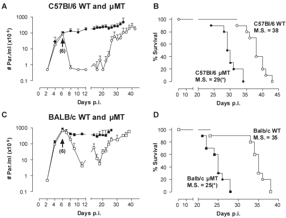
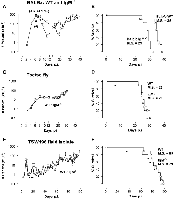
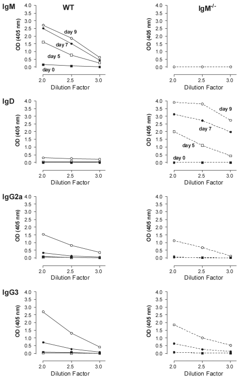
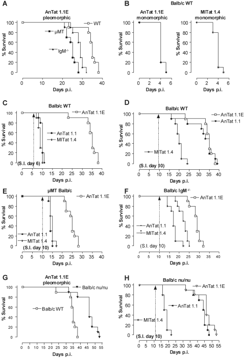
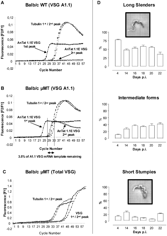
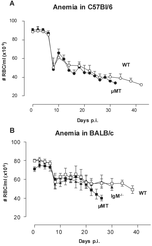
African trypanosomes are extracellular parasitic protozoa, predominantly transmitted by the bite of the haematophagic tsetse fly. The main mechanism considered to mediate parasitemia control in a mammalian host is the continuous interaction between antibodies and the parasite surface, covered by variant-specific surface glycoproteins. Early experimental studies have shown that B-cell responses can be strongly protective but are limited by their VSG-specificity. We have used B-cell (microMT) and IgM-deficient (IgM(-/-)) mice to investigate the role of B-cells and IgM antibodies in parasitemia control and the in vivo induction of trypanosomiasis-associated anemia. These infection studies revealed that that the initial setting of peak levels of parasitemia in Trypanosoma brucei-infected microMT and IgM(-/-) mice occurred independent of the presence of B-cells. However, B-cells helped to periodically reduce circulating parasites levels and were required for long term survival, while IgM antibodies played only a limited role in this process. Infection-associated anemia, hypothesized to be mediated by B-cell responses, was induced during infection in microMT mice as well as in IgM(-/-) mice, and as such occurred independently from the infection-induced host antibody response. Antigenic variation, the main immune evasion mechanism of African trypanosomes, occurred independently from host antibody responses against the parasite's ever-changing antigenic glycoprotein coat. Collectively, these results demonstrated that in murine experimental T. brucei trypanosomiasis, B-cells were crucial for periodic peak parasitemia clearance, whereas parasite-induced IgM antibodies played only a limited role in the outcome of the infection.


Conflict of interest statement

The authors have declared that no competing interests exist.

Figures

Figure 1
Figure 1. The effect of B-cells in trypanosomiasis control of clonal infections.
(A) C57BL/6 WT (○) and µMT (•) mice were infected with a pleomorphic T. brucei AnTat 1.1E clone by intra-peritoneal inoculation of 5000 parasites. Parasitemia was followed by microscopy analysis of tail-cut blood samples. Values are presented as mean±SD of 10 individual mice per group. (B) Mortality of all infected mice was recorded. One representative from 3 experiments is shown. (C,D) The same experiment was performed with BALB/c WT (□) and µMT BALB/c (▪) mice. M.S. = median survival time in days. (*: p<0.0001 compared to WT)
Figure 2
Figure 2. The effect of IgMs in high-, intermediate- or low-virulent infections.
BALB/c WT (□) and IgM−/− (▴) mice were infected with (A/B) 5000 parasites of the pleomorphic AnTat 1.1E clone by intra-peritoneal injection, (C/D) non clonal T. brucei AnTaR 1 parasites through exposure to tsetse fly or (E/F) with 5000 parasites of the low virulent field isolate TSW196 by intra-peritoneal injection. Parasitemia and mortality of infected mice was assessed as in Fig. 1, using 10 mice per experimental group. M.S. = median survival time.
Figure 3
Figure 3. Trypanosome infected IgM−/− mice produce compensatory IgD titers.
Sera were collected from T. brucei AnTat 1.1E infected WT and IgM−/− BALB/c mice, and were analyzed as serial dilutions (log 10) in a solid phase VSG coated ELISA, using isotype-specific antibodies for detection. Sera were collected throughout the first peak and clearance phase from 5 individual mice. Values are presented as means.
Figure 4
Figure 4. The role of IgM in VSG-specific protection.
(A) BALB/c WT (□), IgM−/− (▴) and µMT (▪) mice were infected with 5000 pleomorphic AnTat 1.1E parasites. (B) BALB/c WT mice were infected with 5000 monomorphic AnTat 1.1 (•) or MITat 1.4 (▒) parasites. (C) AnTat 1.1E infected BALB/c WT (□) mice were super-infected (S.I.) on day 6 or (D) on day 10 with 5000 parasites of a homologous, monomorphic AnTat 1.1 (•) or a non-homologous monomorphic, MITat 1.4 (▒) strain. (E) AnTat 1.1E infected BALB/c µMT (□) and (F) BALB/c IgM−/− (□) mice were super-infected with AnTat 1.1 (•) or MITat 1.4 (▒) parasites using the same strategy described above in C–D. (G) BALB/c WT (□) and BALB/c nu/nu (▾) mice were infected with 5000 AnTat 1.1E parasites and (H) were super-infected AnTat 1.1 (•) or MITat 1.4 (▒) parasites using the same strategy described above in C–D. All primary and super-infections were done by intra-peritoneal inoculation of 5000 parasites. Mortality was recorded using 10 mice per experimental group and the results were compared to mice that only received the primary infection. One out of 3 representative experiments is shown.
Figure 5
Figure 5. Quantitative analysis of VSG switching.
Switching of VSG expression was followed by real time RT-PCR, by amplification of peak stage parasite RNA with primers specific for VSG AnTat 1.1E and the housekeeping genes tubulin-zeta (Tubulin). (A) RNA was isolated from WT derived and (B) µMT derived parasites on the first and second peak, respectively occurring on day 6 and 14. Expression of VSG AnTat 1.1E specific RNA on the second peak was calculated and compared to the first peak, after normalization of the first and second peak VSG AnTat 1.1E specific RNA to tubulin-zeta gene expression.. One representative of two experiments are shown. (C) Similarly, total VSG mRNA was amplified from first and second peak µMT derived parasites, using VSG-Uni primers, and results were normalized using Tubulin gene expression. (D) To assess parasite proliferation in µMT mice, the occurrence of dividing ‘long slender’ parasites versus intermediate and non-dividing ‘short stumpy’ parasites was analyzed by light microscopy on day 4 and compared to the chronic infection stage (up to day 22).
Figure 6
Figure 6. Trypanosomiasis-associated anemia is present in µMT mice.
(A) C57BL/6 WT (○) and µMT (•) mice were infected with a pleomorphic T. brucei AnTat 1.1E clone by intra-peritoneal inoculation of 5000 parasites. (B) BALB/c WT (□), µMT (▪) and IgM−/− (▴) mice were infected by intra-peritoneal inoculation of 5000 with the pleomorphic AnTat 1.1E clone. RBC counts were followed by microscopy analysis of tail-cut blood samples. Values are presented as mean±SD of 10 individual mice per group.

References

    1. Vickerman K. Developmental cycles and biology of pathogenic trypanosomes. Br Med Bull. 1985;41:105–114. - PubMed
    1. WHO Control and surveillance of African trypanosomiasis. Report of a WHO Expert Committee. World Health Organ Tech Rep Ser. 1998;881:I–VI. 1–114. - PubMed
    1. Van den Bossche P. Some general aspects of the distribution and epidemiology of bovine trypanosomosis in southern Africa. Int J Parasitol. 2001;31:592–598. - PubMed
    1. van Meirvenne N. Paris ; London: Springer; 1999. Biological diagnosis of human African trypanosomiasis. Progress in human African trypanosomiasis, sleeping sickness. pp. 235–252.
    1. Vanhamme L, Pays E, McCulloch R, Barry JD. An update on antigenic variation in African trypanosomes. Trends Parasitol. 2001;17:338–343. - PubMed

Publication types

MeSH terms