. 2008 Aug 8;4(8):e1000119.
doi: 10.1371/journal.ppat.1000119.

Structure of the head of the Bartonella adhesin BadA

Affiliations

Structure of the head of the Bartonella adhesin BadA

Pawel Szczesny et al. PLoS Pathog. .

Abstract

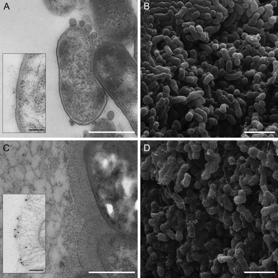
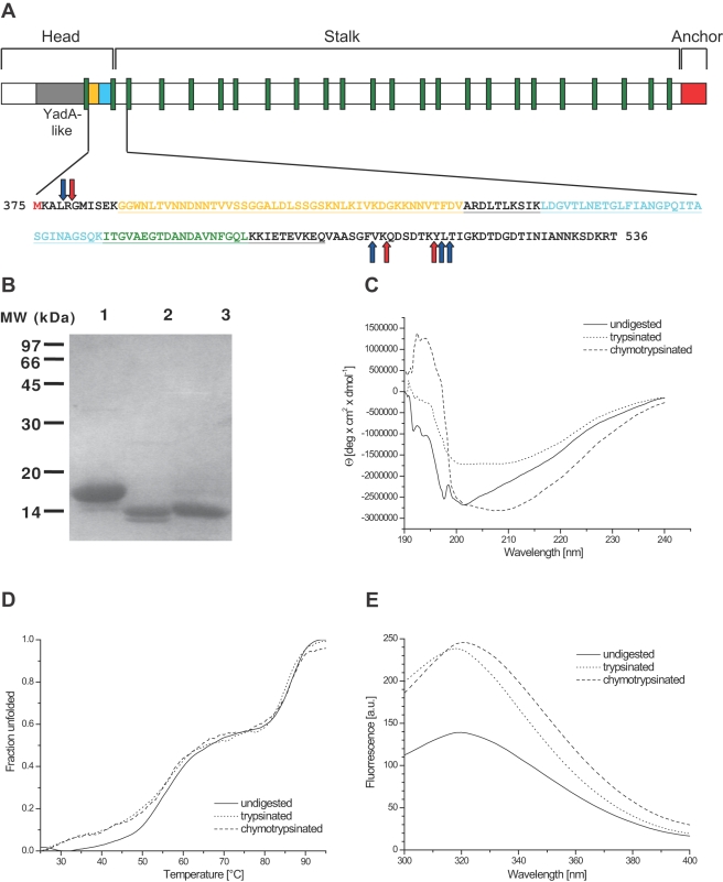
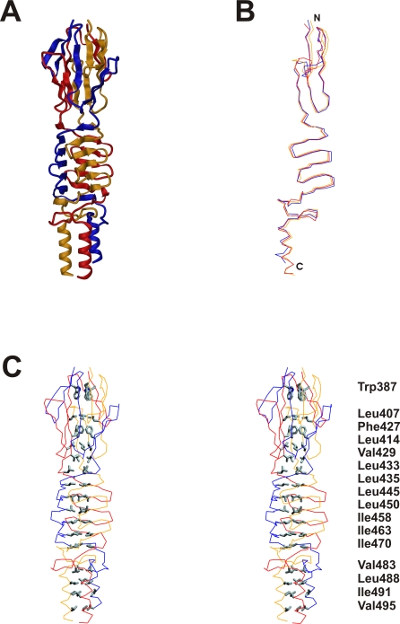
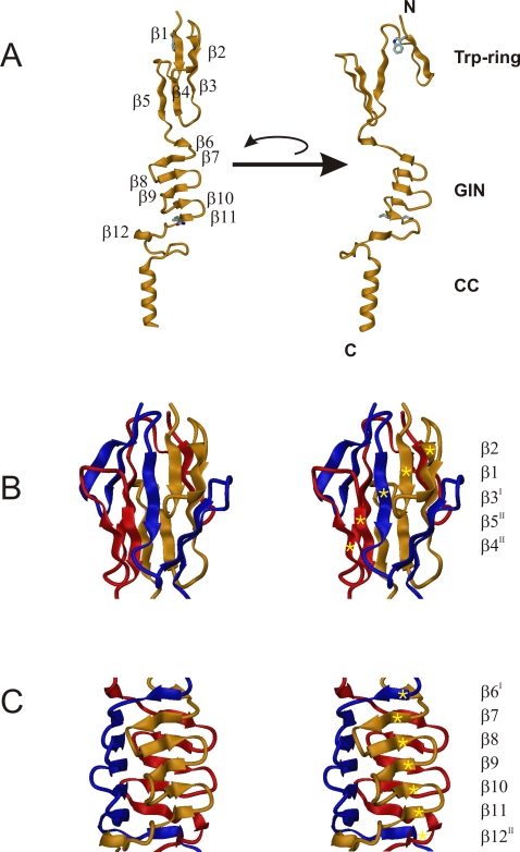
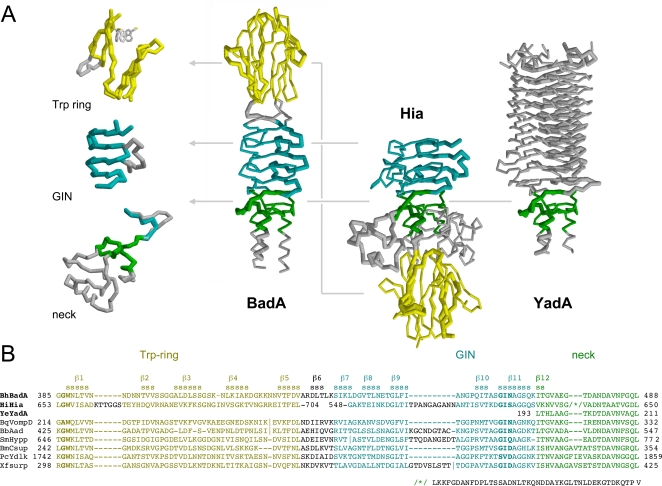
Trimeric autotransporter adhesins (TAAs) are a major class of proteins by which pathogenic proteobacteria adhere to their hosts. Prominent examples include Yersinia YadA, Haemophilus Hia and Hsf, Moraxella UspA1 and A2, and Neisseria NadA. TAAs also occur in symbiotic and environmental species and presumably represent a general solution to the problem of adhesion in proteobacteria. The general structure of TAAs follows a head-stalk-anchor architecture, where the heads are the primary mediators of attachment and autoagglutination. In the major adhesin of Bartonella henselae, BadA, the head consists of three domains, the N-terminal of which shows strong sequence similarity to the head of Yersinia YadA. The two other domains were not recognizably similar to any protein of known structure. We therefore determined their crystal structure to a resolution of 1.1 A. Both domains are beta-prisms, the N-terminal one formed by interleaved, five-stranded beta-meanders parallel to the trimer axis and the C-terminal one by five-stranded beta-meanders orthogonal to the axis. Despite the absence of statistically significant sequence similarity, the two domains are structurally similar to domains from Haemophilus Hia, albeit in permuted order. Thus, the BadA head appears to be a chimera of domains seen in two other TAAs, YadA and Hia, highlighting the combinatorial evolutionary strategy taken by pathogens.


Conflict of interest statement

The authors have declared that no competing interests exist.

Figures

Figure 1
Figure 1. EM pictures of BadA.
Transmission (left) and scanning electron micrographs (right) of Bartonella henselae. Panels A and B show a mutant strain deficient in BadA, panels C and D show the wildtype bacteria grown on blood agar plates. The scale bars are 500 nm in the TEM pictures (A and C) and 2 µm in the SEM pictures (B and D). BadA has a length of 240+/−10 nm. The inserts in panels A and C (scale bar 100 nm) show bacteria after on-section labeling with an antibody raised against the C-terminal head fragment of BadA. The head structure is found exclusively at the tips of the elongated fibers in panel C.
Figure 2
Figure 2. Domain structure of BadA and sequence of the fragment used in this study.
(A) The domain arrangement of BadA, with the YadA-like head in grey, the two domains described in this paper in gold and cyan, respectively, the neck sequences in green and the membrane anchor in red. The lower panel shows the sequence of the fragment used in this study, colored according to the domain arrangement. Red (trypsin) and blue (chymotrypsin) arrows indicate protease cleavage sites; several variants of a protease-resistant 14 kDa core fragment were found by mass spectrometry. Underlined is the part of the sequence that is resolved in the crystal structure, which correlates well with the protease-resistant part of the protein. (B) SDS-PAGE of the 17 kDa fragment before (lane 1) and after trypsin (lane 2) or chymotrypsin (lane 3) treatment. (C) CD spectra of the fragments before and after proteolysis. A higher fraction of random coil signal contributes to the spectrum of the undigested fragment. (D) Heat denaturation of the fragments before and after proteolysis measured at 210 nm wavelength. Unfolding occurs in two steps. (E) Fluorescence spectra of the fragments before and after proteolysis.
Figure 3
Figure 3. Quartenary structure of the BadA head domain.
(A) Structure of the entire BadA head fragment. The three independent protein chains are colored in yellow, red and blue. (B) Superposition of the three individual protein chains. A significant deviation is visible in particular at the N-terminal part of the structure. (C) Stereo representation of the hydrophobic core of the protein, which is built by 16 residues related by threefold symmetry.
Figure 4
Figure 4. Crystal structure of the BadA head domain.
(A) Structure of the monomeric BadA fragment in ribbon representation with the secondary structure elements marked (β1-β12). Two individual orientations rotated around the threefold axis by 90 degrees relative to each other are shown, and the two domains (Trp-ring domain and GIN domain) and the coiled-coil part (CC) are indicated. The conserved residues Trp387 of the Trp-ring and Gly462-Ile463-Asn464 of the GIN domain which determine their nomenclature are shown in stick representation. (B) Structure of the N-terminal Trp-ring domain in stereo representation. The three independent protein chains are color-coded in yellow, red and blue. The three chains form intertwined mixed parallel/anti-parallel β-sheets, one of which is labeled with gold stars. Its β-strand sequence is β2-β1-β3I-β5II-β4II. (C) Structure of the GIN domain in stereo representation, with the individual monomers color-coded in yellow, red and blue. The three interdigitated chains form three β-sheets, one of which is labeled with gold stars. Its β-strand sequence is β6I-β7-β8-β9-β10-β11-β12II. The strand progression of the individual sheets is anti-parallel (β7-β11) while interacting strands from adjacent chains are combined via parallel strand pairing (β6I-β7 and β11-β12II).
Figure 5
Figure 5. Crystal structure of the BadA head domain - omit map.
The |Fobs-Fcalc| electron density around the three Trp387 residues of the Trp-ring domain (calculated after simulated annealing with the Trp sidechains omitted), contoured at 3.5 sigma level.
Figure 6
Figure 6. Network of hydrogen bonds in the necks of BadA, YadA and Hia.
Intramolecular H-bonds are marked as blue lines, intermolecular ones as red lines. Residues involved in intermolecular H-bonds are yellow, residues involved in intramolecular H-bonds are blue, highly conserved hydrophobic core residues that also contribute to the H-bond network are green. Note that all H-bonds involve atoms of the main chains, which explains the low conservation of sidechains between neck sequences. * marks the insertion in the Hia sequence (see text).
Figure 7
Figure 7. Structure comparison of the complete BadA head with YadA from Y. enterolitica and Hia from H. influenzae.
(A) Structures of BadA, Hia and YadA heads with the three domains colored according to the domain annotation from the alignment . The superimpositions of the individual domains from all three proteins are shown in the left panel. Note the different order of domains between Hia and BadA. In the BadA Trp-ring domain, 43 of 45 residues could be superimposed to the equivalent Hia domain with an r.m.s.d. of 2.02 Å, and in the GIN domain, 26 of 30 residues could be superimposed with an r.m.s.d. of 1.58 Å. In the BadA neck region, 19 residues could be superimposed to the YadA neck with an r.m.s.d. of 0.28 Å and to the Hia neck with an r.m.s.d. of 1.32 Å. All r.m.s.d. values refer to the Cα atoms. (B) Sequence alignment of the BadA head with other TAAs. The sequences of Hia and YadA are taken from the published structures; alignments based on these structures were used for homology modeling of the BadA head. The conserved residues that were used to name the domains are marked in bold. Abbreviations used: BhBadA – Bartonella henselae BadA gi|119890727|, HiHia – Haemophilus influenzae Hia gi|21536216|, YeYadA – Yersinia enterocolitica YadA gi|28372996|, BqVompD – Bartonella quintana VompD gi|49473810|, BbAdh – Bartonella bacilliformis adhesin gi|121601790|, SmHypp – Sinorhizobium meliloti hypothetical protein gi|15964211|, BmCsup – Brucella melitensis cell surface protein gi|17988156|, PcYdlk – Psychrobacter cryohalolentis YadA-like protein gi|93006053|, XfSurp – Xylella fastidiosa surface protein gi|15838130|.
Figure 8
Figure 8. Model of the full BadA head.
The head of BadA, comprising the crystal structure of the Trp-ring and GIN domain and models of the YadA-like head and the connecting coiled coil. The structure is heavily intertwined and each chain spirals over 360 degrees around the fiber axis, mostly due to the two neck sequences present.

References

    1. Barocchi MA, Masignani V, Rappuoli R. Opinion: Cell entry machines: a common theme in nature? Nat Rev Microbiol. 2005;3:349–358. - PMC - PubMed
    1. Henderson IR, Navarro-Garcia F, Desvaux M, Fernandez RC, Ala'Aldeen D. Type V protein secretion pathway: the autotransporter story. Microbiol Mol Biol Rev. 2004;68:692–744. - PMC - PubMed
    1. Linke D, Riess T, Autenrieth IB, Lupas A, Kempf VA. Trimeric autotransporter adhesins: variable structure, common function. Trends Microbiol. 2006;14:264–270. - PubMed
    1. Hoiczyk E, Roggenkamp A, Reichenbecher M, Lupas A, Heesemann J. Structure and sequence analysis of Yersinia YadA and Moraxella UspAs reveal a novel class of adhesins. EMBO J. 2000;19:5989–5999. - PMC - PubMed
    1. Cover TL, Aber RC. Yersinia enterocolitica. N Engl J Med. 1989;321:16–24. - PubMed

Publication types

MeSH terms

Substances