. 2008 Nov 1;17(21):3399-410.
doi: 10.1093/hmg/ddn234. Epub 2008 Aug 8.

SMN complex localizes to the sarcomeric Z-disc and is a proteolytic target of calpain

Affiliations

SMN complex localizes to the sarcomeric Z-disc and is a proteolytic target of calpain

Michael P Walker et al. Hum Mol Genet. .

Abstract

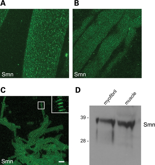
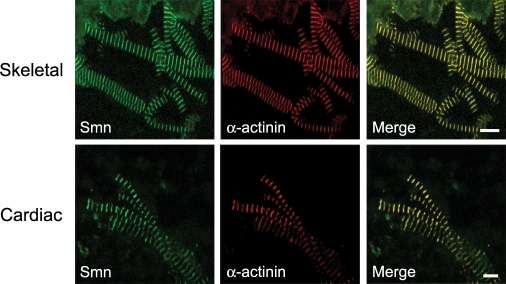
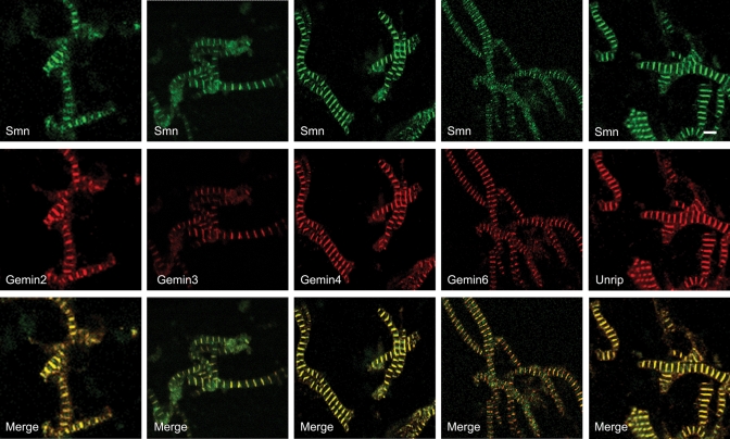
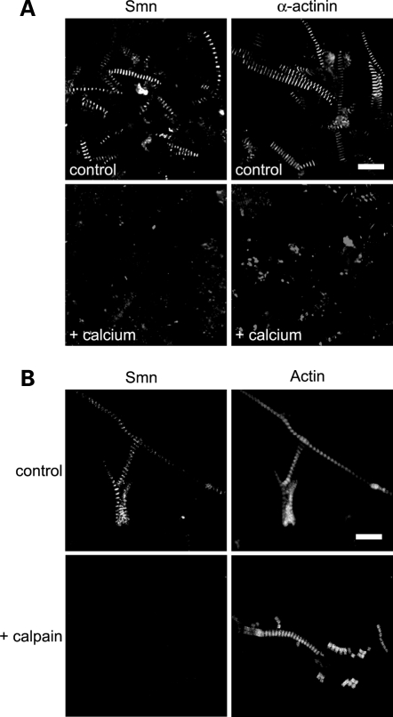
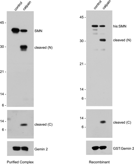
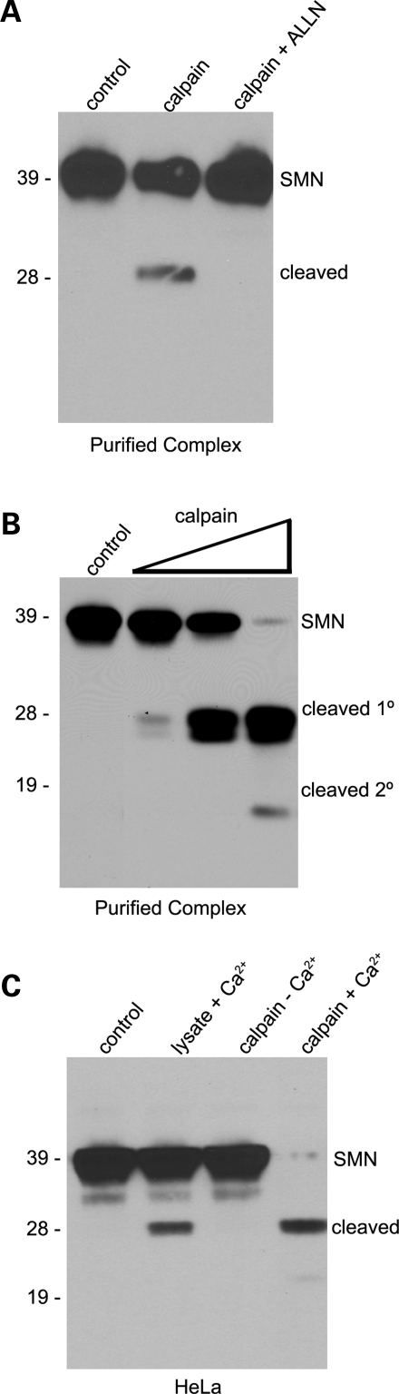
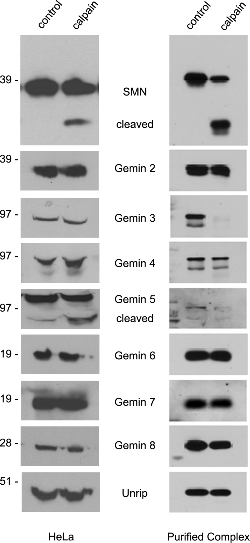
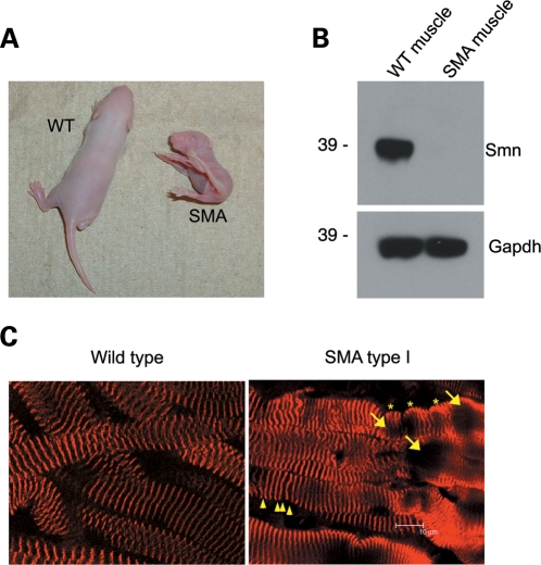
Spinal muscular atrophy (SMA) is a recessive neuromuscular disease caused by mutations in the human survival motor neuron 1 (SMN1) gene. The human SMN protein is part of a large macromolecular complex involved in the biogenesis of small ribonucleoproteins. Previously, we showed that SMN is a sarcomeric protein in flies and mice. In this report, we show that the entire mouse Smn complex localizes to the sarcomeric Z-disc. Smn colocalizes with alpha-actinin, a Z-disc marker protein, in both skeletal and cardiac myofibrils. Furthermore, this localization is both calcium- and calpain-dependent. Calpains are known to release proteins from various regions of the sarcomere as a part of the normal functioning of the muscle; however, this removal can be either direct or indirect. Using mammalian cell lysates, purified native SMN complexes, as well as recombinant SMN protein, we show that SMN is a direct target of calpain cleavage. Finally, myofibers from a mouse model of severe SMA, but not controls, display morphological defects that are consistent with a Z-disc deficiency. These results support the view that the SMN complex performs a muscle-specific function at the Z-discs.


Figures

Figure 1.
Figure 1.
Smn localizes to skeletal myofibrils. Mouse muscle fibers and purified myofibrils were subjected to immunofluorescence imaging (average projection of a Z-stack of 13 focal planes, imaged at 0.5 µm per section) and western blotting. Antibodies against Smn (mAb Clone 8) were used to stain muscle fibers (A, B) and purified myofibrils (C); scale bar represents 10 µm. The presence of Smn was verified by western blot of whole muscle lysate and purified myofibrils (D).
Figure 2.
Figure 2.
Smn localizes to the Z-discs of striated muscle. Purified myofibrils from skeletal and cardiac muscle were co-stained with antibodies against Smn and the Z-disc protein α-actinin; the scale bar for skeletal myofibrils is 10 and 5 µm for cardiac myofibrils.
Figure 3.
Figure 3.
Smn complex is present at Z-Discs. Purified mouse hindlimb skeletal myofibrils were co-stained with antibodies against Smn and Gemins 2–4, 6 and Unrip. Because the anti-Gemin 3 antibody (mAb 12H12) does not cross-react with mouse, but does cross-react with the hamster protein (http://www.abcam.com/index.html?datasheet=10305), purified myofibrils from hamster hindlimb were used for the Gemin 3 panel. Scale bar represents 5 µm.
Figure 4.
Figure 4.
U snRNPs do not localize to myofibrils. Purified skeletal myofibrils were co-stained with an antibody against the U2-specific protein, U2B′′, an antibody specific to the U snRNP Sm core (mAb Y12), an Sm-core protein, SmB (mAb 12F5) and an antibody specific to the U snRNP 5′-TMG cap, TMG (mAb K121) (AC). The myofibrils were counter stained with phaloidin conjugated with FITC. A negative control antibody (neurofilament-L) was also used on purified myofibrils (D); scale bars represent 10 µm.
Figure 5.
Figure 5.
Calpain activity removes Smn from myofibrils. Increased endogenous calpain activity was indirectly assessed by incubating skeletal muscle tissue either in a buffer with 10 mm calcium, which activates calpain, or in a buffer lacking calcium; purified myofibrils were either stained with antibodies against Smn or α-actinin as a positive control (A). Exogenous calpain 1 was incubated with purified skeletal myofibrils (controls were incubated without added calpain) and co-stained with antibodies against Smn and conjugated phalloidin (B). Scale bars 10 µm.
Figure 6.
Figure 6.
Calpain directly cleaves SMN in vitro. To verify that calpain cleaves SMN directly, purified SMN complexes or recombinant His-SMN/GST-Gemin 2 heterodimers were incubated with 0.06 and 0.15 U of calpain, respectively. Antibodies recognizing the N- (Clone 8) and C-terminal (mAb 9F2) domains of SMN were used to visualize the two fragments of SMN from the purified SMN complexes. His-probe reagent was used to visualize recombinant His-SMN.
Figure 7.
Figure 7.
Calpain inhibition, titration and endogenous activation. Purified native SMN complexes were treated with calpain (0.05 U) or with calpain and ALLN inhibitor. The cleavage of SMN was compared with an undigested control lane (A). Increasing amounts of calpain (0.003 to 3.0 U) were incubated with purified SMN complexes and the cleavage profile of SMN was analyzed (B). Western blotting was performed on HeLa lysates using anti-SMN (Clone 8) antibodies. In lanes 2 and 4, 1 mm calcium was added to the lysates. In lanes 3 and 4, 1.5 U of calpain were added. Lysates were incubated for 15 min. at 30°C and SMN cleavage profiles were compared with the control (lane 1) (C).
Figure 8.
Figure 8.
Calpain cleaves a subset of the Gemins within the SMN complex in vitro and in vivo. HeLa lysate and purified SMN complexes were treated with exogenous calpain, 1.4 and 0.06 U, respectively. Western blotting was performed with antibodies to each member of the SMN complex. Note control and calpain lanes in the Gemin 5 and Gemin 3 panels.
Figure 9.
Figure 9.
SMA type 1 mice have aberrant myofibrils. Muscle fibers from SMA and wild-type littermates (A, B) were fixed and then stained with an antibody against α-actinin (average projection of a Z-stack of 18 focal planes at 0.5 µm per section) and compared (C). See text for details.

Similar articles

Cited by

References

    1. Wirth B., Brichta L., Hahnen E. Spinal muscular atrophy: from gene to therapy. Semin. Pediatr. Neurol. 2006;13:121–131. - PubMed
    1. Pellizzoni L. Chaperoning ribonucleoprotein biogenesis in health and disease. EMBO Rep. 2007;8:340–345. - PMC - PubMed
    1. Kolb S.J., Battle D.J., Dreyfuss G. Molecular functions of the SMN complex. J. Child Neurol. 2007;22:990–994. - PubMed
    1. Sumner C.J. Molecular mechanisms of spinal muscular atrophy. J. Child Neurol. 2007;22:979–989. - PubMed
    1. Eggert C., Chari A., Laggerbauer B., Fischer U. Spinal muscular atrophy: the RNP connection. Trends Mol. Med. 2006;12:113–121. - PubMed

Publication types

MeSH terms