. 2008;59(11):3187-201.
doi: 10.1093/jxb/ern175.

Pollination in Nicotiana alata stimulates synthesis and transfer to the stigmatic surface of NaStEP, a vacuolar Kunitz proteinase inhibitor homologue

Affiliations

Pollination in Nicotiana alata stimulates synthesis and transfer to the stigmatic surface of NaStEP, a vacuolar Kunitz proteinase inhibitor homologue

Grethel Yanet Busot et al. J Exp Bot. 2008.

Abstract

After landing on a wet stigma, pollen grains hydrate and germination generally occurs. However, there is no certainty of the pollen tube growth through the style to reach the ovary. The pistil is a gatekeeper that evolved in many species to recognize and reject the self-pollen, avoiding endogamy and encouraging cross-pollination. However, recognition is a complex process, and specific factors are needed. Here the isolation and characterization of a stigma-specific protein from N. alata, NaStEP (N. alata Stigma Expressed Protein), that is homologous to Kunitz-type proteinase inhibitors, are reported. Activity gel assays showed that NaStEP is not a functional serine proteinase inhibitor. Immunohistochemical and protein blot analyses revealed that NaStEP is detectable in stigmas of self-incompatible (SI) species N. alata, N. forgetiana, and N. bonariensis, but not in self-compatible (SC) species N. tabacum, N. plumbaginifolia, N. benthamiana, N. longiflora, and N. glauca. NaStEP contains the vacuolar targeting sequence NPIVL, and immunocytochemistry experiments showed vacuolar localization in unpollinated stigmas. After self-pollination or pollination with pollen from the SC species N. tabacum or N. plumbaginifolia, NaStEP was also found in the stigmatic exudate. The synthesis and presence in the stigmatic exudate of this protein was strongly induced in N. alata following incompatible pollination with N. tabacum pollen. The transfer of NaStEP to the stigmatic exudate was accompanied by perforation of the stigmatic cell wall, which appeared to release the vacuolar contents to the apoplastic space. The increase in NaStEP synthesis after pollination and its presence in the stigmatic exudates suggest that this protein may play a role in the early pollen-stigma interactions that regulate pollen tube growth in Nicotiana.


Figures

Fig. 1.
Fig. 1.
Species-specific and developmental expression of NaStEP and NaSoEP. (a) Detection of NaStEP in different genetic backgrounds. Total RNA (10 μg) from mature pistils of SI N. alata (Na) and SC N. plumbaginifolia (Np), N. longiflora (Nl), and N. tabacum (Nt) was blotted and hybridized with 32P-labelled NaStEP. (b) NaStEP expression in developing anthers and pistils of N. alata SC10SC10. Total RNA (10 μg) was loaded in each lane, blotted, and probed with 32P-labelled NaStEP. (c) NaSoEP expression in developing anthers and pistils of N. alata SC10SC10. Total RNA (10 μg) was loaded in each lane, blotted, and probed with 32P-labelled NaSoEP. (d) Densitometric analysis of NaStEP and NaSoEP expression during the development of styles plus stigmas. Numbers 1–5 represents the developmental stages of stigmas plus styles: 1, 0.5–1.0 cm; 2, 1–2 cm; 3, 2–3.5 cm; 4, 3.5–6 cm; and 5, 6 cm. (e) NaStEP and NaSoEP expression in non-sexual organs of N. alata. Total RNA (10 μg) was loaded in each lane, blotted, and probed with a 32P-labelled NaStEP (upper) or NaSoEP (lower). The same amount of stigma RNA was loaded as hybridization control. To ascertain equal RNA loading, blots were stained with methylene blue (lower sections of a–c and e).
Fig. 2.
Fig. 2.
Alignment of the amino acid sequences of the cDNA encoding NaStEP and NaSoEP proteins. The black boxes under the alignment indicate the degree of sequence consensus. Arrowheads indicate the potential signal peptide cleavage sites. Cysteine residues are marked with an asterisk. Putative glycosylation sites are single underlined. Presumed vacuolar sorting signals are boxed. The antigenic region is double underlined. A dash in the sequence indicates a gap introduced to maintain a good alignment.
Fig. 3.
Fig. 3.
Immunoanalysis of NaStEP in N. alata organs. Equal amounts of total protein (10 μg) of roots, stems, leaves, sepals, petals, pistils, styles, stigmas, ovaries, and anthers were separated by SDS–PAGE. (a) Proteins transferred onto nitrocellulose and immunostained with anti-NaStEP antibody. (b) Coomassie blue-stained proteins.
Fig. 4.
Fig. 4.
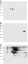
Two-dimensional gel analysis of NaStEP in stigmatic extracts. Total protein (50 μg) from matures styles plus stigmas of SI N. alata SA2SA2 were fractionated by two-dimensional electrophoresis. (a) Proteins transferred onto nitrocellulose and immunodetected with anti-NaStEP antibody. (b) Proteins blotted and immunodetected with the anti-SA2-RNase antibody. (c) Mature proteins silver stained.
Fig. 5.
Fig. 5.
Native NaStEP from N. alata stigmas and serine proteinase inhibitor activity assay. Purified HPLC fractions (29–36) of NaStEP were resolved by SDS–PAGE. (a) Proteins present in the HPLC fractions were transferred onto nitrocellulose and immunostained with the anti-NaStEP antibody. (b) Silver-stained proteins. (c) Inhibitory activity gel assay against trypsin. A 3 μg aliquot of the soybean trypsin inhibitor (STI) was run as a positive control. (d) A silver-stained replicate of the inhibitory activity assay gel.
Fig. 6.
Fig. 6.
Immunohistochemical localization of NaStEP in stigmatic tissues of SC N. alata Breakthrough. (a) Stigma and style treated with pre-immune serum. (b) Stigma and style treated with anti-NaStEP antibody (green). (c) Stigma and style treated with anti-HT-B antibody (magenta). (d) and (e) Diagrams of stigma plus style of N. alata and a transverse section of a stigma. The arrow and square show zones where images were taken. (f) Stigma cross-sections showing NaStEP (magenta) in papillary cells (red arrow), secretory cells (yellow arrow), basal cells (blue arrow), and in a portion of the upper transmitting tract. (g) High magnification view of stigmatic secretory cells shows that NaStEP is in small bodies (blue arrows) and in the proximity of the cell wall (green arrows). All figures represent merges of immunostained and phase contrast images of stigmas plus styles. CW, cell wall; V, vacuole; TT, transmitting tissue; UTT, upper transmitting track; C, cortex; PC, papillae cell. Bars, 50 μm for (a), (b), (c) and 20 μm for (f) and 5 μm for (g).
Fig. 7.
Fig. 7.
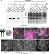
NaStEP-like proteins are expressed in stigmas of SI Nicotiana species but no in SC species. (a) Total proteins (10 μg) from stigmas and styles of SI N. bonariensis, SI N. forgetiana, SI N. alata SA2SA2, SI N. alata S105SC10, SI N. alata S105S105, SC N. alata S105SA2, SC N. alata Breakthrough, SI Hybrid 1 (SC N. plumbaginifolia×SI N. alata S105S105), SI Hybrid 2 (SC N. plumbaginifolia×SI N. alata SC10SC10), SC N. plumbaginifolia, SC N. tabacum, SC N. longiflora, and SC N. glauca were separated by SDS–PAGE and either Coomassie blue stained (left) or blotted to nitrocellulose for immunostaining with anti-NaStEP antibody (right). (b) Cross-sections of N. alata Breakthrough, SI N. alata SC10SC10, SI N. forgetiana, SC N. tabacum, SC N. plumbaginifolia, SC N. glauca, and SC N. benthamiana stigmas immunostained with anti-NaStEP. I and II: diagrams of a stigma plus style of N. alata and a transverse section of a stigma. The arrow and square show zones where images were taken. UTT, upper transmitting tissue; SC, secretory cells; PC, papillae cell. Bars, 50 mm.
Fig. 8.
Fig. 8.
NaStEP is discharged from the vacuole to the stigmatic exudate upon pollination. Unpollinated stigma from SC N. alata cv Breakthrough showing NaStEP localization in vacuoles and osmiophilic bodies. (a) Cross-section of mature papillae surrounded by copious exudate containing secretory droplets. Arrows show cell wall interruptions. (b) A papillary cell with a large vacuole and osmiophilic bodies. (c) Anti-NaStEP labelling of vacuolar electron-dense bodies. Arrows show immunogold secondary antibody labelling. Nicotiana alata cv Breakthrough stigma 10 h after pollination with N. tabacum pollen. (d) Degenerating papillary cells (arrow), showing abundant exudate and a disordered cytoplasm with abnormal organelles and disintegrated vacuoles. (e) Vacuole showing poor anti-NaStEP labelling of the osmiophilic bodies (arrows). (f) A close-up of the labelling shown in (e) (black arrows) showing anti-NaStEP labelling (white arrows) in smaller osmiophilic bodies. Stigmatic exudate of N. alata cv Breakthrough. (g) and (h) Diagrams of a stigma plus style of N. alata and a transverse section of a stigma. The arrow and square show zones where images were taken. (i) Unpollinated stigmas. (j) At 10 h after pollination with N. tabacum pollen. (k) At 10 h after pollination with N. plumbaginifolia pollen. (l) At 10 h after self-pollination. N, nucleus; m, mitochondria; c, chloroplast; v, vacuole; osb, osmiophilic bodies; ex, exudate. Bars, 500 nm for (a), 1 μm for (b) and (d), 240 nm for (c), 500 nm for (e), and 200 nm for (i)–(l).
Fig. 9.
Fig. 9.
Pollination induces synthesis and release of NaStEP into the N. alata stigmatic exudate. Total protein from exudate of unpollinated and pollinated stigmas of N. alata cv Breakthrough were sequentially extracted, separated by SDS–PAGE, and blotted to nitrocellulose for immunostaining with anti-NaStEP (top) or silver nitrate stained (bottom). Equal volumes (30 μl) were loaded in each lane. (a) Total protein from exudate of unpollinated stigmas at anthesis and at 48 h old. (b) Total proteins from exudate after 5, 12, and 24 h of self-pollination. (c) Total proteins from exudate after 5, 12, and 24 h of pollination with N. tabacum pollen. (d), (e), and (f) Densitometric analysis of NaStEP expression and its presence in the stigmatic exudate of N. alata before and after pollination with self- and N. tabacum pollen. L, low-salt wash; H, high-salt wash; T, proteins remaining in the stigma after washes.

Similar articles

Cited by

References

    1. Altschul SF, Madden TL, Schaffer AA, Zhang J, Zhang Z, Miller W, Lipman DJ. Gapped BLAST and PSI-BLAST: a new generation of protein database search programs. Nucleic Acids Research. 1997;25:3389–3402. - PMC - PubMed
    1. Atkinson AH, Heath RL, Simpson RJ, Clarke AE, Anderson MA. Proteinase inhibitors in Nicotiana alata stigmas are derived from a precursor protein which is processed into five homologous inhibitors. The Plant Cell. 1993;5:203–213. - PMC - PubMed
    1. Beecher B, McClure BA. Effects of RNases on rejection of pollen from Nicotiana tabacum and N. plumbaginifolia. Sexual Plant Reproduction. 2001;14:69–76.
    1. Bendtsen JD, Nielsen H, von Heijne G, Brunak S. Improved prediction of signal peptides: SignalP 3.0. Journal of Molecular Biology. 2004;340:783–795. - PubMed
    1. Bradford MM. A rapid and sensitive method for the quantitation of microgram quantities of protein using the principle of protein–dye binding. Analytical Biochemistry. 1976;72:248–254. - PubMed

Publication types