. 2008 Nov 15;95(10):4597-612.
doi: 10.1529/biophysj.108.128728. Epub 2008 Aug 8.

Calmodulin mediates differential sensitivity of CaMKII and calcineurin to local Ca2+ in cardiac myocytes

Affiliations

Calmodulin mediates differential sensitivity of CaMKII and calcineurin to local Ca2+ in cardiac myocytes

Jeffrey J Saucerman et al. Biophys J. .

Abstract

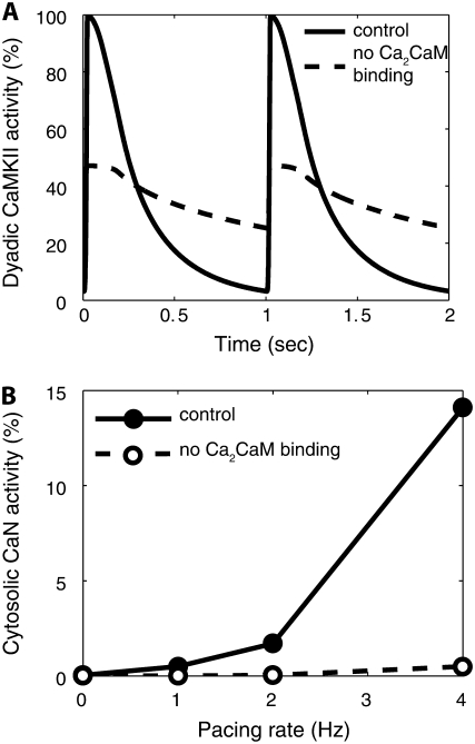
Calmodulin (CaM) mediates Ca-dependent regulation of numerous pathways in the heart, including CaM-dependent kinase (CaMKII) and calcineurin (CaN), yet the local Ca(2+) signals responsible for their selective activation are unclear. To assess when and where CaM, CaMKII, and CaN may be activated in the cardiac myocyte, we integrated new mechanistic computational models of CaM, CaMKII, and CaN with the Shannon-Bers model of excitation-contraction coupling in the rabbit ventricular myocyte. These models are validated with independent in vitro data. In the intact myocyte, model simulations predict that CaM is highly activated in the dyadic cleft during each beat, but not appreciably in the cytosol. CaMKII-delta(C) was almost insensitive to cytosolic Ca due to relatively low CaM affinity. Dyadic cleft CaMKII exhibits dynamic frequency-dependent responses to Ca, yet autophosphorylates only when local phosphatases are suppressed. In contrast, dyadic cleft CaN in beating myocytes is predicted to be constitutively active, whereas the extremely high affinity of CaN for CaM allows gradual integration of small cytosolic CaM signals. Reversing CaM affinities for CaMKII and CaN also reverses their characteristic local responses. Deactivation of both CaMKII and CaN seems dominated by Ca dissociation from the complex (versus Ca-CaM dissociation from the target). In summary, the different affinities of CaM for CaMKII and CaN determine their sensitivity to local Ca signals in cardiac myocytes.


Figures

FIGURE 1
FIGURE 1
Model of calmodulin (CaM)-dependent signaling in cardiac myocytes. (A) Compartmental model schematic of cardiac myocyte EC coupling (7) incorporating CaM, CaMKII, and CaN signaling in thedyadic cleft and cytosol, as described in Methods. (B) Reaction map for cooperative Ca binding of 2 Ca to CaM sequentially to the C-terminal and then N-terminal EF hands, along with binding of CaM “buffers”. (C) Probabilistic model of CaMKIIδ subunit switching between inactive (Pi), inactive Ca2CaM-bound (Pb2), active Ca4CaM-bound (Pb), Thr287-autophosphorylated with Ca4CaM trapped (Pt), and Thr287-autophosphorylated but CaM-autonomous (Pa) or Ca2CaM-bound (Pt2) states. (D) Reaction map for reversible binding of CaM, Ca2CaM, and Ca4CaM to CaN.
FIGURE 2
FIGURE 2
In vitro validation of model components. (A) Model-predicted Ca binding to CaM at 1 mM or 6 mM [Mg2+] and 100 mM [K+] (experimental data from Stemmer and Klee (11)). (B) Predicted [Ca] versus CaN activity for 0.03 and 0.3 μM [CaM, (experimental data from Stemmer and Klee (11)). (C and D) Predicted [CaM] versus CaM-dependent CaMKIIδ activity (C) and CaMKIIδ-Thr287 activity (D) (independent experimental data from Gaertner et al. (6)). (E) Predicted [Ca] versus CaM-dependent CaMKIIδ activity, compared with independent CaMKIIα data from Bradshaw et al. (20). (F) Predicted Thr287P-dependent CaMKIIδ activity for 0 or 2.5 μM [PP1] (independent CaMKIIα data from Bradshaw et al. (20)).
FIGURE 3
FIGURE 3
Model of CaM buffering. (A) Predicted kinetics of CaM/buffer interaction in permeabilized cardiac myocytes, with slower dissociation at high [Ca] (experimental data from Wu and Bers (23)). (B) As [Ca] increases, buffered CaM is predicted to switch from CaMB (solid line) to Ca2CaMB (dashed line) to Ca4CaMB (dotted line).
FIGURE 4
FIGURE 4
Local CaM dynamics in a model of a beating cardiac myocyte. (A) Ca2CaM and Ca4CaM are strongly activated during each beat in the dyadic cleft. (B) Only a small fraction of CaM binds Ca in the cytosol with 1 Hz pacing. Ca dissociates very rapidly from Ca4CaM and more slowly from Ca2CaM. (C) When pacing rate is increased from 1 to 4 Hz, Ca4CaM still does not accumulate in the dyadic cleft. (D) Ca4CaM only slightly accumulates activity in the cytosol. (E) Subsarcolemmal Ca4CaM signals are predicted to lie between dyadic and cytosolic [Ca4CaM].
FIGURE 5
FIGURE 5
Predicted local CaMKII dynamics in beating cardiac myocytes. (A) Dyadic-cleft CaMKII activity increases significantly from 1 (solid line) to 4 Hz pacing (dashed line). (B) Dyadic-cleft CaMKII-Thr287P autophosphorylation is minimal under normal conditions, unless PP1 tethering to RyR is disrupted, as seen in heart failure (37,39). (C) Cytosolic CaMKII activity remains very low at 1 and 4 Hz.
FIGURE 6
FIGURE 6
Predicted local calcineurin dynamics in beating cardiac myocytes. (A) Even at low heart rates, any CaN in the dyadic cleft remains fully locked in an activated state due to its slow dissociation of Ca4CaM. (B) When pacing is increased from 0.5 to 4 Hz, cytosolic CaN activity slowly accumulates activity from previous beats with τ ∼ 15 min. (C) Cytosolic CaN is fairly sensitive to pacing rate, integrating dynamic Ca signals with activity somewhat higher than in response to a constant signal fixed at the mean cytosolic [Ca].
FIGURE 7
FIGURE 7
Reversing CaM affinity switches characteristic local activation patterns of CaMKII and CaN. Increasing CaMKII's affinity for Ca4CaM to that of CaN (28 pM) enhances cytosolic activity (A) yet locks dyadic-cleft CaMKII in a fully activated state. Lowering CaN's affinity for Ca4CaM to that of CaMKII (33.5 nM) allows appreciable beat-to-beat variation of CaN activity in the dyadic cleft (B) yet substantially reduces cytosolic CaN activity (C).
FIGURE 8
FIGURE 8
Role of CaM buffering in modulating CaMKII and CaN activation. Increasing CaM availability by eliminating CaM sarcolemmal and cytosolic CaM buffers (BTOT-SL = 0, BTOT-CYT = 0) (dashed lines) is insufficient to substantially activate cytosolic CaMKII (A), but significantly enhances cytosolic CaN activity in response to pacing (B).
FIGURE 9
FIGURE 9
Role of Ca2CaM binding in modulating CaMKII and CaN activation. (A) Eliminating binding reactions between Ca2CaM and CaMKII reduced peak dyadic-cleft CaMKII activity and slowed inactivation, leaving mean activity relatively unaffected at 1 Hz. (B) Eliminating Ca2CaM binding substantially suppressed cytosolic CaN activity.

Similar articles

Cited by

References

    1. Bush, E. W., D. B. Hood, P. J. Papst, J. A. Chapo, W. Minobe, M. R. Bristow, E. N. Olson, and T. A. McKinsey. 2006. Canonical transient receptor potential channels promote cardiomyocyte hypertrophy through activation of calcineurin signaling. J. Biol. Chem. 281:33487–33496. - PubMed
    1. Molkentin, J. D. 2006. Dichotomy of Ca2+ in the heart: contraction versus intracellular signaling. J. Clin. Invest. 116:623–626. - PMC - PubMed
    1. Persechini, A., and P. M. Stemmer. 2002. Calmodulin is a limiting factor in the cell. Trends Cardiovasc. Med. 12:32–37. - PubMed
    1. Berridge, M. J. 2006. Calcium microdomains: organization and function. Cell Calcium. 40:405–412. - PubMed
    1. Quintana, A. R., D. Wang, J. E. Forbes, and M. N. Waxham. 2005. Kinetics of calmodulin binding to calcineurin. Biochem. Biophys. Res. Commun. 334:674–680. - PubMed

Publication types

LinkOut - more resources