. 2008 Oct;190(20):6749-57.
doi: 10.1128/JB.00589-08. Epub 2008 Aug 8.

Polar localization and compartmentalization of ClpP proteases during growth and sporulation in Bacillus subtilis

Affiliations

Polar localization and compartmentalization of ClpP proteases during growth and sporulation in Bacillus subtilis

James Kain et al. J Bacteriol. 2008 Oct.

Abstract

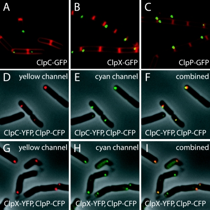
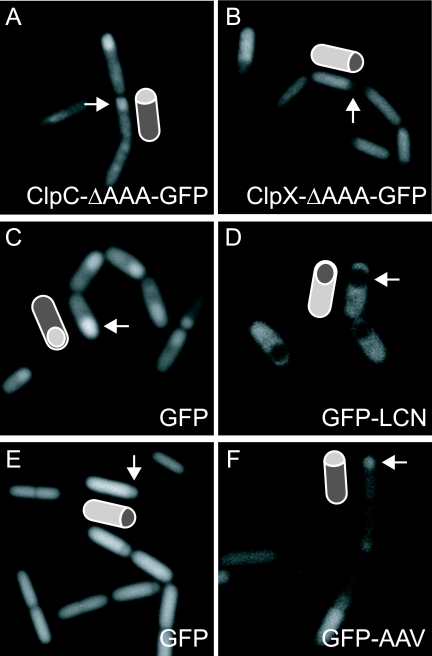
Spatial control of proteolysis is emerging as a common feature of regulatory networks in bacteria. In the spore-forming bacterium Bacillus subtilis, the peptidase ClpP can associate with any of three ATPases: ClpC, ClpE, and ClpX. Here, we report that ClpCP, ClpEP, and ClpXP localize in foci often near the poles of growing cells and that ClpP and the ATPase are each capable of polar localization independently of the other component. A region of ClpC containing an AAA domain was necessary and sufficient for polar localization. We also report that ClpCP and ClpXP proteases differentially localize to the forespore and mother cell compartments of the sporangium during spore formation. Moreover, model substrates for each protease created by appending recognition sequences for ClpCP or ClpXP to the green fluorescent protein were preferentially eliminated from the forespore or the mother cell, respectively. Biased accumulation of ClpCP in the forespore may contribute to the cell-specific activation of the transcription factor sigma(F) by preferential ClpCP-dependent degradation of the anti-sigma(F) factor SpoIIAB.


Figures

FIG. 1.
FIG. 1.
Subcellular localization of ClpC, ClpX, and ClpP. The coding sequence for GFP was fused to the coding sequence for ClpC (A; BJK166), ClpX (B; BJK210), or ClpP (C; BJK207), and images were captured from cells harboring the fusions by using fluorescence microscopy during exponential growth in LB medium. Membranes were stained with the dye FM4-64. Panels D to F show cells from a strain (BJK499) harboring both clpC-yfp and clpP-cfp. The cells were grown on LB plates and imaged by fluorescence microscopy in the yellow channel (false-colored red) (D), the cyan channel (false-colored green) (E), and merged (F). The yellow and cyan fluorescence signals were overlaid with the phase-contrast image. Panels G to I show cells from a strain (BJK485) harboring both clpX-yfp and clpP-cfp. Cells were taken from plates and imaged with fluorescence microscopy in the yellow channel (false-colored red) (G), the cyan channel (false-colored green) (H), and merged (I). The yellow and cyan fluorescence signals were overlaid with the phase-contrast image.
FIG. 2.
FIG. 2.
Polar localizations of ClpP and its ATPases are not interdependent. Panel A shows the effects of the indicated clp mutations on the localization of the indicated Clp-GFP fusions. The strains used were BJK243 (a), BJK235 (b), BJK200 (c), BJK242 (d), BJK234 (e), and BJK370 (f). Panel B shows the localization of the indicated Clp-GFP fusions in the presence of no mutation or the indicated multiple mutations. The strains used were BJK285 (a), BJK465 (b), and BJK474 (c). Panel C shows the localization of fusions of GFP to the indicated B. subtilis Clp proteins produced in E. coli. The gene fusions for the proteins were expressed from an IPTG-inducible promoter, and the images were captured 30 min after the addition of IPTG (1 mM final concentration) during exponential-phase growth in CH medium. Membranes were stained with the dye FM4-64 and overlaid with the GFP signal. The strains used were BJK476 (a), BJK477 (b), and BJK505 (c).
FIG. 3.
FIG. 3.
An AAA-containing domain of ClpC is sufficient for polar focus formation. Panel A shows the anatomy of Clp ATPases and the end points of the indicated truncations of ClpC. The ATPases have one or two nucleotide-binding domains designated AAA1 or AAA2, with AAA1 domains more similar to each other than to AAA2 domains. Additional domains are labeled as follows: N, Clp N domain; L, UVR linker domain; Z, zinc binding domain. Horizontal lines indicate truncations from the N-terminal or C-terminal direction, with short vertical lines designating the end points. Panel B shows the localization pattern of GFP fused to the indicated truncations of ClpC. The gene fusions were expressed from either the native promoter (a to c) or from IPTG-inducible promoters (d to f), and images were captured 30 min after the addition of IPTG (1 mM final concentration) to cells in the exponential phase of growth in CH medium. Fluorescence from staining with the dye FM4-64 was overlaid with the signal from GFP. The cells were B. subtilis, except for those in image f, in which the indicated ClpC truncation was artificially produced in E. coli by use of an IPTG-inducible promoter. The strains were BJK166 (a), BJK552 (b), BJK317 (c), BJK359 (d), BJK506 (e), and BJK504 (f).
FIG. 4.
FIG. 4.
Subcellular localization of ClpC, ClpX, and ClpP during sporulation. Panel A shows the subcellular localization of ClpX-GFP (a; BJK210) and ClpP-GFP (b; BJK207) 3 h after the induction of sporulation by resuspension. Arrows highlight large foci in the mother cell. Arrowheads highlight smaller foci frequently found on the forespore. Panel B shows a time course of ClpC-GFP (BJK166) during sporulation. Cells were imaged at 0.5 (a), 2 (b), 3 (c), and 5 (d) hours after the induction of sporulation by resuspension. In image b, the arrow highlights a spirallike focus pattern in the mother cell, and the arrowhead highlights foci tracking with the engulfing membranes. In image c, arrows highlight a diffuse GFP signal in the mother cell while arrowheads highlight foci associated with the forespore. In image d, arrowheads highlight delocalized foci in the forespore. All images were captured by fluorescence microscopy. Membranes were stained with the dye FM4-64 and overlaid with the GFP signal. Also shown in the panels are interpretive cartoons.
FIG. 5.
FIG. 5.
ClpCP and ClpXP exhibit compartment-specific biases in localization and activity during sporulation. Panels A and B show that the N-terminal portion of ClpC or ClpX fused to GFP accumulates in the forespore or the mother cell, respectively. The strains used were BJK315 (A) and BJK311 (B). Images were taken using fluorescence microscopy 2 h after initiation of sporulation by resuspension. Panels C and D show the pattern of accumulation of GFP (C; BJK510) or GFP-LCN (D; BJK456) produced from copies of the corresponding coding sequences that had been introduced into the chromosome just downstream of the native locus for clpC. Images were captured at hour 3 after resuspension in SM medium. Panels E and F show the pattern of accumulation of GFP (E; BJK458) or GFP-LCN (F; BJK456) produced under the control of an IPTG-inducible promoter. Images were captured at hour 1.5 after resuspension. Also shown in the panels are interpretative cartoons, with arrows identifying the forespores.
FIG. 6.
FIG. 6.
Model for a self-reinforcing cycle that contributes to the activation of σF. Panel A summarizes the interrelationships among the anti-σF anti-sigma factor SpoIIAB (AB), the anti-anti-sigma factor SpoIIAA (AA), the inactive, phosphorylated form of SpoIIAA (AA-P), the SpoIIAA-P phosphatase SpoIIE (E), and ClpCP. AB is both an anti-sigma factor and a protein kinase that phosphorylates AA. Unphosphorylated AA can both displace σF from the AB-σF complex and become trapped in an inactive complex with AB (AA-AB). Notice that removal of AB by degradation increases the relative amount of AA, leading to increased disruption of σF-AB and hence increased levels of free and active σF. Panel B depicts a hypothesized self-reinforcing cycle in which σF drives additional synthesis of ClpC, which, in turn, further reduces the levels of the AB anti-sigma factor, leading to yet higher levels of free and active σF.

Similar articles

Cited by

References

    1. Alba, B. M., J. A. Leeds, C. Onufryk, C. Z. Lu, and C. A. Gross. 2002. DegS and YaeL participate sequentially in the cleavage of RseA to activate the sigma(E)-dependent extracytoplasmic stress response. Genes Dev. 162156-2168. - PMC - PubMed
    1. Andersen, J. B., C. Sternberg, L. K. Poulsen, S. P. Bjorn, M. Givskov, and S. Molin. 1998. New unstable variants of green fluorescent protein for studies of transient gene expression in bacteria. Appl. Environ. Microbiol. 642240-2246. - PMC - PubMed
    1. Ben-Yehuda, S., M. Fujita, X. S. Liu, B. Gorbatyuk, D. Skoko, J. Yan, J. F. Marko, J. S. Liu, P. Eichenberger, D. Z. Rudner, and R. Losick. 2005. Defining a centromere-like element in Bacillus subtilis by identifying the binding sites for the chromosome-anchoring protein RacA. Mol. Cell 17773-782. - PubMed
    1. Bernhardt, T. G., and P. A. de Boer. 2005. SlmA, a nucleoid-associated, FtsZ binding protein required for blocking septal ring assembly over chromosomes in E. coli. Mol. Cell 18555-564. - PMC - PubMed
    1. Derre, I., G. Rapoport, K. Devine, M. Rose, and T. Msadek. 1999. ClpE, a novel type of HSP100 ATPase, is part of the CtsR heat shock regulon of Bacillus subtilis. Mol. Microbiol. 32581-593. - PubMed

Publication types

MeSH terms

LinkOut - more resources