

The two-component system PhoPR of Clostridium acetobutylicum is involved in phosphate-dependent gene regulation
- PMID: 18689481
- PMCID: PMC2566200
- DOI: 10.1128/JB.00574-08
The two-component system PhoPR of Clostridium acetobutylicum is involved in phosphate-dependent gene regulation
Abstract
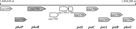
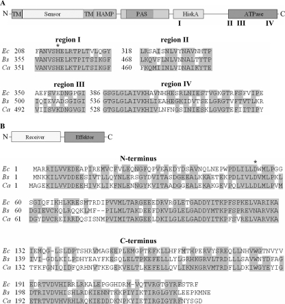
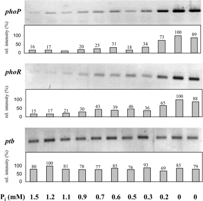
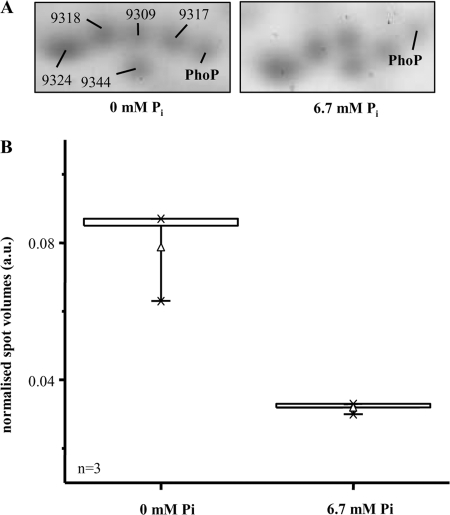
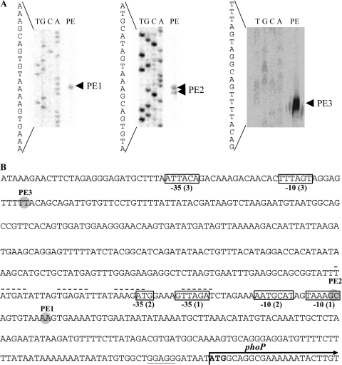
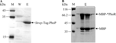
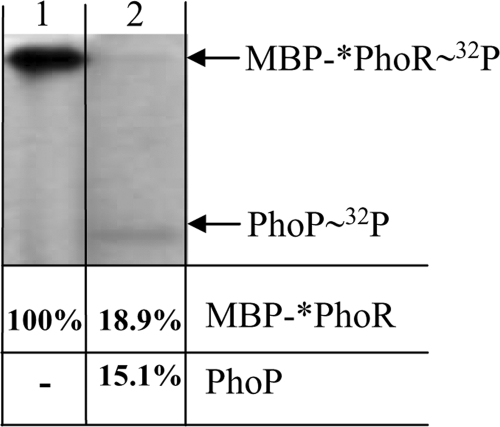
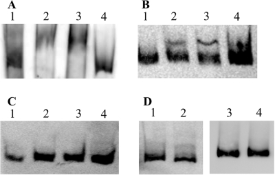
The phoPR gene locus of Clostridium acetobutylicum ATCC 824 comprises two genes, phoP and phoR. Deduced proteins are predicted to represent a response regulator and sensor kinase of a phosphate-dependent two-component regulatory system. We analyzed the expression patterns of phoPR in P(i)-limited chemostat cultures and in response to P(i) pulses. A basic transcription level under high-phosphate conditions was shown, and a significant increase in mRNA transcript levels was found when external P(i) concentrations dropped below 0.3 mM. In two-dimensional gel electrophoresis experiments, a 2.5-fold increase in PhoP was observed under P(i)-limiting growth conditions compared to growth with an excess of P(i). At least three different transcription start points for phoP were determined by primer extension analyses. Proteins PhoP and an N-terminally truncated *PhoR were individually expressed heterologously in Escherichia coli and purified. Autophosphorylation of *PhoR and phosphorylation of PhoP were shown in vitro. Electromobility shift assays proved that there was a specific binding of PhoP to the promoter region of the phosphate-regulated pst operon of C. acetobutylicum.
Figures
Similar articles
-
Functional characterization of PhoPR two component system and its implication in regulating phosphate homeostasis in Bacillus anthracis.Biochim Biophys Acta Gen Subj. 2017 Jan;1861(1 Pt A):2956-2970. doi: 10.1016/j.bbagen.2016.09.022. Epub 2016 Sep 23. Biochim Biophys Acta Gen Subj. 2017. PMID: 27667172
-
Transcription of the pst operon of Clostridium acetobutylicum is dependent on phosphate concentration and pH.J Bacteriol. 2006 Aug;188(15):5469-78. doi: 10.1128/JB.00491-06. J Bacteriol. 2006. PMID: 16855236 Free PMC article.
-
Transcriptional regulation of the phoPR operon in Bacillus subtilis.J Bacteriol. 2004 Feb;186(4):1182-90. doi: 10.1128/JB.186.4.1182-1190.2004. J Bacteriol. 2004. PMID: 14762014 Free PMC article.
-
Transcriptomic studies of phosphate control of primary and secondary metabolism in Streptomyces coelicolor.Appl Microbiol Biotechnol. 2012 Jul;95(1):61-75. doi: 10.1007/s00253-012-4129-6. Epub 2012 May 24. Appl Microbiol Biotechnol. 2012. PMID: 22622839 Review.
-
Activation of the PhoPR-Mediated Response to Phosphate Limitation Is Regulated by Wall Teichoic Acid Metabolism in Bacillus subtilis.Front Microbiol. 2018 Nov 6;9:2678. doi: 10.3389/fmicb.2018.02678. eCollection 2018. Front Microbiol. 2018. PMID: 30459743 Free PMC article. Review.
Cited by
-
Integrative modelling of pH-dependent enzyme activity and transcriptomic regulation of the acetone-butanol-ethanol fermentation of Clostridium acetobutylicum in continuous culture.Microb Biotechnol. 2013 Sep;6(5):526-39. doi: 10.1111/1751-7915.12033. Epub 2013 Jan 21. Microb Biotechnol. 2013. PMID: 23332010 Free PMC article.
-
A systems biology approach to investigate the effect of pH-induced gene regulation on solvent production by Clostridium acetobutylicum in continuous culture.BMC Syst Biol. 2011 Jan 19;5:10. doi: 10.1186/1752-0509-5-10. BMC Syst Biol. 2011. PMID: 21247470 Free PMC article.
-
Exploring the rearrangement of sensory intelligence in proteobacteria: insight of Pho regulon.World J Microbiol Biotechnol. 2018 Nov 9;34(11):172. doi: 10.1007/s11274-018-2551-3. World J Microbiol Biotechnol. 2018. PMID: 30413888
-
Mathematical modelling of clostridial acetone-butanol-ethanol fermentation.Appl Microbiol Biotechnol. 2017 Mar;101(6):2251-2271. doi: 10.1007/s00253-017-8137-4. Epub 2017 Feb 16. Appl Microbiol Biotechnol. 2017. PMID: 28210797 Free PMC article. Review.
-
A proteomic and transcriptional view of acidogenic and solventogenic steady-state cells of Clostridium acetobutylicum in a chemostat culture.Appl Microbiol Biotechnol. 2010 Aug;87(6):2209-26. doi: 10.1007/s00253-010-2741-x. Epub 2010 Jul 9. Appl Microbiol Biotechnol. 2010. PMID: 20617312 Free PMC article.
References
-
- Aguena, M., E. Yagil, and B. Spira. 2002. Transcriptional analysis of the pst operon of Escherichia coli. Mol. Genet. Genomics 268518-524. - PubMed
Publication types
MeSH terms
Substances
LinkOut - more resources
Full Text Sources
Molecular Biology Databases
Research Materials
Miscellaneous
