. 2008 Oct;74(19):5925-33.
doi: 10.1128/AEM.00318-08. Epub 2008 Aug 8.

DNA fragmentation in microorganisms assessed in situ

Affiliations

DNA fragmentation in microorganisms assessed in situ

José Luis Fernández et al. Appl Environ Microbiol. 2008 Oct.

Abstract

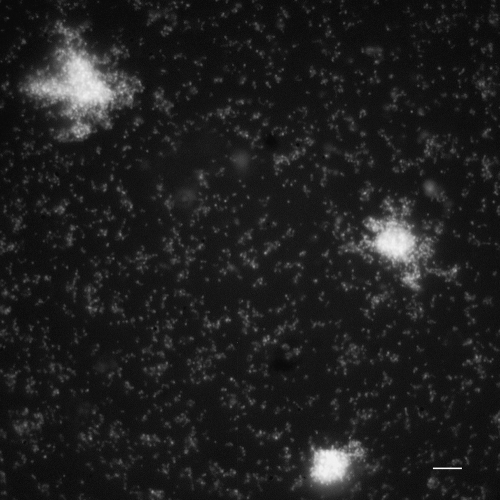
Chromosomal DNA fragmentation may be a direct or indirect outcome of cell death. Unlike DNA fragmentation in higher eukaryotic cells, DNA fragmentation in microorganisms is rarely studied. We report an adaptation of a diffusion-based assay, developed as a kit, which allows for simple and rapid discrimination of bacteria with fragmented DNA. Intact cells were embedded in an agarose microgel on a slide, incubated in a lysis buffer to partially remove the cell walls, membranes, and proteins, and then stained with a DNA fluorochrome, SYBR Gold. Identifying cells with fragmented DNA uses peripheral diffusion of DNA fragments. Cells without DNA fragmentation show only limited spreading of DNA fiber loops. These results have been seen in several gram-negative and gram-positive bacteria, as well as in yeasts. Detection of DNA fragmentation was confirmed by fluoroquinolone treatment and by DNA breakage detection-fluorescence in situ hybridization. Proteus mirabilis with spontaneously fragmented DNA during exponential and stationary growth or Escherichia coli with DNA damaged after exposure to hydrogen peroxide or antibiotics, such as ciprofloxacin or ampicillin, was clearly detected. Similarly, fragmented DNA was detected in Saccharomyces cerevisiae after amphotericin B treatment. Our assay may be useful for the simple and rapid evaluation of DNA damage and repair as well as cell death, either spontaneous or induced by exogenous stimuli, including antimicrobial agents or environmental conditions.


Figures

FIG. 1.
FIG. 1.
Images after application of the Micro-Halomax kit to E. coli cultures. Cells were embedded in an agarose microgel, lysed, and stained with SYBR Gold. (a) Nucleoids from control untreated cells, showing the spread of DNA loops from a central core. One has highly diffused DNA spots (asterisk). (b) Detailed image of an undamaged nucleoid, showing an uneven microgranular surface and multibranched appearance. (c) Ciprofloxacin treatment at the MIC dose, 0.012 μg/ml, for 40 min resulted in DNA fragments. (d) These DNA fragments increased after a treatment of 5 μg/ml. Nucleoids show a big halo of diffusion of DNA spots, similar to those in the image marked by an asterisk in panel a. Bar: 5 μm in panels a, c, and d; 2.5 μm in panel b.
FIG. 2.
FIG. 2.
DBD-FISH-detected DNA breaks in nucleoids from control cultures of E. coli. Cells in an agarose microgel were lysed and treated with an alkaline unwinding treatment to transform DNA breaks into restricted ssDNA motifs detected by hybridization with a whole-genome probe, Cy-3 labeled (red). The central nucleoid with diffused DNA spots appears intensely labeled, confirming the presence of spontaneous massive DNA breaks (asterisk). Nucleoids without fragmented DNA were visible with DAPI (blue). Bar: 5 μm.
FIG. 3.
FIG. 3.
Images after the application of Micro-Halomax to cultures of P. mirabilis (a) and C. albicans (b), processed as indicated in Materials and Methods. Nucleoids without DNA fragmentation released DNA loops around a central core from the residual cell. Otherwise, nucleoids with fragmented DNA were clearly identified by a big halo of DNA spots (asterisks). Bar: 5 μm.
FIG. 4.
FIG. 4.
Kinetics of the frequency of P. mirabilis cells with fragmented DNA and with a PI-permeable membrane. Identification of bacteria with fragmented DNA was performed using the Micro-Halomax kit, as indicated in Materials and Methods. OD600, optical density at 600 nm.
FIG. 5.
FIG. 5.
Exponentially growing cultures from E. coli control cells (a) and from those exposed to 10 mM hydrogen peroxide for 10 min (b), evaluated with the diffusion-based assay using the Micro-Halomax kit. (a) Bacterial nucleoids from control cultures only show spreading of DNA loops. Some nucleoids had a big halo of diffused DNA spots, as indicated in the image (asterisk). (b) All nucleoids observed after H2O2 treatment reveal a halo of DNA spots. In stationary-phase cultures, untreated cells (c) showed similar images to those in panel a, with a higher proportion of background nucleoids with diffused DNA spots (asterisks), whereas those treated with H2O2 (d) were similar to those in panel b. Bar: 5 μm.
FIG. 6.
FIG. 6.
E. coli cultures processed with the Micro-Halomax kit after ampicillin treatment, 300 μg/ml, for 24 h. Nucleoids from residual cells appear more relaxed, accompanied by a dense background of DNA fragments. Bar: 5 μm.
FIG. 7.
FIG. 7.
S. cerevisiae cells with fragmented DNA (right scale), a PI-permeable membrane, and viability (left scale) after incubation for 24 h with increasing doses of amphotericin B.

References

    1. Aersten, A., and C. W. Michiels. 2004. Stress and how bacteria cope with death and survival. Crit. Rev. Microbiol. 30:263-273. - PubMed
    1. Ahnström, G. 1988. Techniques to measure DNA strand breaks in cells: a review. Int. J. Radiat. Biol. 54:695-707. - PubMed
    1. Ahnström, G., and K. Erixon. 1973. Radiation-induced strand breakage in DNA from mammalian cells. Strand separation in alkaline solution. Int. J. Radiat. Biol. 23:285-289. - PubMed
    1. Baginski, M., J. Czub, and K. Sternal. 2006. Interaction of amphotericin B and its selected derivatives with membranes: molecular modeling studies. Chem. Rec. 6:320-332. - PubMed
    1. Bayles, K. W. 2000. The bactericidal action of penicillin: new clues to an unsolved mystery. Trends Microbiol. 8:274-278. - PubMed

Publication types

LinkOut - more resources