Comparative Study
. 2008 Sep 12;103(6):580-90.
doi: 10.1161/CIRCRESAHA.108.171835. Epub 2008 Aug 8.

Perinatal loss of Nkx2-5 results in rapid conduction and contraction defects

Affiliations
Comparative Study

Perinatal loss of Nkx2-5 results in rapid conduction and contraction defects

Laura E Briggs et al. Circ Res. .

Abstract

Homeobox transcription factor Nkx2-5, highly expressed in heart, is a critical factor during early embryonic cardiac development. In this study, using tamoxifen-inducible Nkx2-5 knockout mice, we demonstrate the role of Nkx2-5 in conduction and contraction in neonates within 4 days after perinatal tamoxifen injection. Conduction defect was accompanied by reduction in ventricular expression of the cardiac voltage-gated Na+ channel pore-forming alpha-subunit (Na(v)1.5-alpha), the largest ion channel in the heart responsive for rapid depolarization of the action potential, which leads to increased intracellular Ca2+ for contraction (conduction-contraction coupling). In addition, expression of ryanodine receptor 2, through which Ca2+ is released from sarcoplasmic reticulum, was substantially reduced in Nkx2-5 knockout mice. These results indicate that Nkx2-5 function is critical not only during cardiac development but also in perinatal hearts, by regulating expression of several important gene products involved in conduction and contraction.


Figures
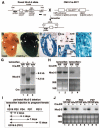
Figure 1
Figure 1
Schematics of generation of tamoxifen-inducible Nkx2-5 knockout mice. A, Homozygous mice for floxed-Nkx2-5 were bred with heterozygous Cre-ER™ transgenic mice. Subsequent matings between offspring generated Nkx2-5flox/flox/Tg-Cre-ER™ (flox/flox/Cre), Nkx2-5wild/wild/Tg-Cre-ER™ (wild/wild/Cre), and Nkx2-5flox/flox (flox/flox). A 8.3-kb XbaI-digested genomic DNA fragments hybridized with the 5′ probe were reduced to 4.3 kb after tamoxifen-induced Cre-recombinase activity. Nkx2-5 gene has 2 coding exons. B through H, Recombination efficiency of Tg-Cre-ER™ in R26R reporter mice. B, Whole-mount staining for β-galactosidase with or without Cre-ER™ transgene. Frozen heart sections of R26R/Tg-Cre-ER™ stained for β-galactosidase. C through F, Four chamber saggital section (C), acetylcholine esterase staining of AV node (D) and adjacent tissue section including AV node (E) and left ventricle (F). Scale bars: 500 μm (B and C); 200 μm (D and E); and 20 μm (F). G, Southern blotting confirmed 8.3-kb genomic DNA fragment isolated from hearts of flox/flox/Cre mice without tamoxifen injection (lane 1) and 4.3 kb after tamoxifen injection (lane 2). Cre-mediated recombination was absent after tamoxifen injection in homozygous wild type carrying the Cre-ER™ transgene (lane 3). H, Northern blotting demonstrated that Nkx2-5 mRNA remained unchanged without tamoxifen injections, but was below detection level with tamoxifen injection (lane 2 vs 4). I, Experimental time course study of Nkx2-5 perinatal knockout. J, Northern blotting confirmed near complete loss of Nkx2-5 mRNA from PD2 to PD7 in flox/flox/Cre mice (lanes 2, 5, and 8). HD indicates homeodomain; fl, flox; w, wild; RA, right atrium; LA, left atrium; RV, right ventricle; LV, left ventricle; TV, tricuspid valve; MV, mitral valve; AV, atrioventricular.
Figure 2
Figure 2
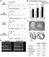
Progressive AV block, prolongation of QRS duration, and heart enlargement in Nkx2-5 knockout mice. A, Surface ECG recordings (lead I) from PD4 and PD12 flox/flox mice showed normal sinus rhythm, but PD4 flox/flox/Cre mice showed PR-prolongation and left bundle branch block, and flox/flox/Cre mice at PD12 showed independent P and QRS waves (complete AV block) with wide QRS complex (see the Table). B, Heart weight/body weight (mg/g) of flox/flox, flox/flox/Cre and wild/wild/Cre mice with tamoxifen injections (mean±SE). C, Hearts dissected from PD12 flox/flox and flox/flox/Cre mice demonstrate dilation of atria and ventricles in Nkx2-5 knockout mice. Hematoxylin/eosin-stained heart sections are shown. Scale bars=1 mm. D, Representative imaging of M-mode ultrasound biomicroscope. Echocardiographic indices of control and Nkx2-5 knockout mice around PD12 (mean±SE) are shown. SCL indicates sinus cycle length (beat-to-beat heart rate); LVDd, left ventricular diastolic dimension; LVDs, left ventricular systolic dimension; HR, heart rate; %FS, percentage of left ventricular fractional shortening.
Figure 3
Figure 3
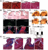
Nkx2-5 protein expression in contractile myocardium and AV node. A, Coimmunostaining of Nkx2-5 (green) and sarcomeric actinin (red) demonstrate reduced expression of Nkx2-5 proteins in ventricles dissected from Nkx2-5 knockout mice at PD4 (left images). Heart sections from PD12 mice stained with Masson’s trichrome show no apparent interstitial fibrosis in Nkx2-5 knockout mice (right images). B, Tissue sections of PD4 hearts demonstrate AV nodes positive for acetylcholine esterase in flox/flox and flox/flox/Cre mice (top images, arrowheads). Adjacent tissue sections including AV node are positive for Nkx2-5 (green) in flox/flox mice but not in flox/flox/Cre mice (middle images). Merged images of Nkx2-5 (green), sarcomeric actinin (red), and nuclear stainings (blue) are shown (bottom images). Scale bars=100 μm/L. C, Whole-mount acetylcholine esterase staining demonstrates AV nodes (brown) both in flox/flox and flox/flox/Cre mice at PD4 and PD12. Scale bars=1 mm. D, Surface area positive for acetylcholine esterase compared between flox/flox and flox/flox/Cre mice at PD4 and PD12 (mean±SE). *P<0.01. E, No fibrosis was observed in Nkx2-5 knockout AV node by Masson’s trichrome staining at PD12. AV nodal area is marked with white dots. Scales are indicated. A indicates atrium; V, ventricle; RA, right atrium; LA, left atrium; RV, right ventricle; TV, tricuspid valve; MV, mitral valve; Ao, aorta; IVS, interventricular septum.
Figure 4
Figure 4
Reduced expression of ion channels in Nkx2-5 knockout hearts. A, Real-time RT-PCR shows fold difference (Nkx2-5 vs control) of mRNA of T-type Ca channels (α1G and α1H), Nav1.5(α), and RyR2 at PD2 and 7. B, Northern blotting reveals KcneI/minK and HOP/HOD mRNA reduction in Nkx2-5 knockout hearts. Of note, HOP/HOD is reported as a direct target of Nkx2-5., HOP/HOD indicates homeodomain only protein. See supplemental Tables I and II.
Figure 5
Figure 5
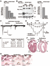
Reduced expression of Nav1.5 α-subunit and reduced INa in Nkx2-5 knockout ventricles. A, Real-time RT-PCR demonstrates progressive reduction of Nav1.5(α) mRNA at PD2, PD4, PD7, and PD12. B, Western blotting demonstrates progressive reduction of Nav1.5(α) protein at PD4 and 12 (lanes 1 vs 2 and 3 vs 4). Fold difference compared to control hearts normalized to GAPDH is shown in right image. Nkx2-5 proteins (arrowheads) are below the level of detection in flox/flox/Cre mice at PD4 and PD12 (arrowheads). C, Representative INa trace recordings from cardiac myocytes obtained from flox/flox (left) and flox/flox/Cre mice (right) using 10 mmol/L Na+ as charge carrier. Myocytes were maintained at -110 mV holding potential with stimulation to -80 to +10 mV at 10-mV steps for 100 ms at 0.1 Hz. D, Current-voltage relation plot for INa in flox/flox (n=6) and flox/flox/Cre (n=7) myocytes, total 7 to 8 days after tamoxifen injections. INa are normalized by cell capacitance and presented as mean±SE. E, Table of kinetic values with maximal current density obtained at -30-mV stimulation. V50, obtained from steady-state activation analysis is the voltage potential determined to elicit the half-maximal INa; KmV is the slope factor; and Vrev is the reversal potential for INa. F, Immunostaining of Nav1.5(α) demonstrates reduced Nav1.5(α) protein in ventricles from Nkx2-5 knockout mice (right images, arrowheads) but preserved expression in atria at PD12 (right images, arrows). Scale bar=1 mm. G, Immunostaining of Nav1.5(α) protein (top) and acetylcholine esterase staining in adjacent tissue sections (bottom). In flox/flox mice, part of AV node is positive for Nav1.5(α) (left images). In flox/flox/Cre mice, serial sections including AV node demonstrate Nav1.5(α) staining absent in the section shown in middle images but is present at a limited part of AV node in the section shown in right images. Scale bars=500 μm. *P<0.01.
Figure 6
Figure 6
Proximal promoter of Scn5a gene contains Nkx2-5 responsive elements. A, Schematics of 2 Nkx2-5 consensus binding sites in the Scn5a promoter at -628 and -590 bp. Scn5a promoter has CpG islands, but not a TATA box. Scn5a(-736 to +119) (top) and mutated Scn5a(-736 to +119) (bottom), and corresponding relative luciferase reporter activities with pcDNA3-Nkx2-5 cotransfection normalized to β-galactosidase activity and pcDNA empty vector with the value in promoter-less luciferase reporter defined as 1 (means ± SE). ANOVA demonstrated significant difference between 2 groups (F=22.1, P=0.0003). *P=0.0003 by Bonferroni’s post hoc test. B, Schematics of conserved genomic regions among mouse, rat, and human Scn5a gene approximately -27, -17, -10, -8, and -2 kb upstream of the transcriptional start site. Six luciferase reporter constructs and corresponding relative luciferase reporter activities with pcDNA3-Nkx2-5 cotransfection normalized to β-galactosidase activity and pcDNA empty vector with the value in promoter-less luciferase reporter defined as 1 (means±SE). ANOVA demonstrated no significant difference among groups (F=1.08, P=0.3966).
Figure 7
Figure 7
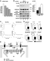
Reduced contractility accompanied by Ca2+-handling defects in cardiomyocytes isolated from Nkx2-5 knockout heart. A, Real-time RT-PCR demonstrates progressive reduction of RyR2 mRNA at PD2, PD4, PD7, and PD12. B, Western blotting demonstrates marked reduction of RyR2 proteins and modest reduction of SERCA2a and phospholamban (PLN) (pentamer and monomer) in heart lysates obtained from Nkx2-5 knockout mice compared to control mice at PD12. Fold difference compared to control hearts normalized with GAPDH is shown at right (mean±SE, n=2). Note, because of different molecular sizes of RyR2 (≈560 kDa), phospholamban monomer (≈5 kDa) and GAPDH (≈30 kDa), equal amounts of protein were loaded on separate gels. Signal intensities in Western blotting were normalized to those of GAPDH. C, Representative tracings of a single contraction (top) and simultaneous Ca2+ transients (bottom) in an isolated cardiomyocyte from control flox/flox (left) or Nkx2-5 knockout (flox/flox/Cre) mice (right). D, Summarized data (mean±SE) obtained from multiple cardiomyocytes demonstrate that Nkx2-5 knockout cardiomyocytes show decreased percentage fractional shortening, +dL/dT (speed of contraction), and -dL/dT (speed of relaxation), Ca2+ amplitude, Ca2+-releasing velocity, and Ca2+ decay constant. *P<0.01. E, Simplified Na+ and Ca2+ transport through sarcolemma and SR in cardiomyocytes (modified from a previously published figure41). Model for the altered excitation-contraction coupling in Nkx2-5 knockout cardiomyocytes; reduced Na+ inward currents through Nav1.5, reduced Ca2+ inward currents possibly through T-type Ca2+ channels and reverse mode of sodium-calcium exchanger (increased Na+ entry and Ca2+ exit) may reduce Ca2+ inflow into cytosol (dotted lines). In addition, Ca2+ release from SR to cytosol, as well as Ca2+ uptake to SR, is reduced by reduction of RyR2 and SERCA2a in Nkx2-5 knockout hearts (dotted lines). Overall, altered cytosolic Ca2+ handling would decrease cardiac contraction.

Similar articles

Cited by

References

    1. Bers DM. Cardiac excitation-contraction coupling. Nature. 2002;415:198–205. - PubMed
    1. Marban E. Cardiac channelopathies. Nature. 2002;415:213–218. - PubMed
    1. Mangoni ME, Couette B, Marger L, Bourinet E, Striessnig J, Nargeot J. Voltage-dependent calcium channels and cardiac pacemaker activity: from ionic currents to genes. Prog Biophys Mol Biol. 2006;90:38–63. - PubMed
    1. Haufe V, Chamberland C, Dumaine R. The promiscuous nature of the cardiac sodium current. J Mol Cell Cardiol. 2007;42:469–477. - PubMed
    1. Abriel H. Roles and regulation of the cardiac sodium channel Na(v) 1.5: recent insights from experimental studies. Cardiovasc Res. 2007;76:381–389. - PubMed

Publication types

MeSH terms