. 2008 Aug 19;105(33):11748-53.
doi: 10.1073/pnas.0805852105. Epub 2008 Aug 8.

Modulation of actin structure and function by phosphorylation of Tyr-53 and profilin binding

Affiliations

Modulation of actin structure and function by phosphorylation of Tyr-53 and profilin binding

Kyuwon Baek et al. Proc Natl Acad Sci U S A. .

Abstract

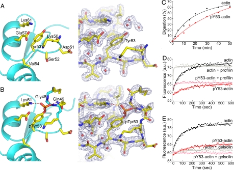
On starvation, Dictyostelium cells aggregate to form multicellular fruiting bodies containing spores that germinate when transferred to nutrient-rich medium. This developmental cycle correlates with the extent of actin phosphorylation at Tyr-53 (pY53-actin), which is low in vegetative cells but high in viable mature spores. Here we describe high-resolution crystal structures of pY53-actin and unphosphorylated actin in complexes with gelsolin segment 1 and profilin. In the structure of pY53-actin, the phosphate group on Tyr-53 makes hydrogen-bonding interactions with residues of the DNase I-binding loop (D-loop) of actin, resulting in a more stable conformation of the D-loop than in the unphosphorylated structures. A more rigidly folded D-loop may explain some of the previously described properties of pY53-actin, including its increased critical concentration for polymerization, reduced rates of nucleation and pointed end elongation, and weak affinity for DNase I. We show here that phosphorylation of Tyr-53 inhibits subtilisin cleavage of the D-loop and reduces the rate of nucleotide exchange on actin. The structure of profilin-Dictyostelium-actin is strikingly similar to previously determined structures of profilin-beta-actin and profilin-alpha-actin. By comparing this representative set of profilin-actin structures with other structures of actin, we highlight the effects of profilin on the actin conformation. In the profilin-actin complexes, subdomains 1 and 3 of actin close around profilin, producing a 4.7 degrees rotation of the two major domains of actin relative to each other. As a result, the nucleotide cleft becomes moderately more open in the profilin-actin complex, probably explaining the stimulation of nucleotide exchange on actin by profilin.


Conflict of interest statement

The authors declare no conflict of interest.

Figures

Fig. 1.
Fig. 1.
Conformational change in actin subdomain 2 on phosphorylation of Tyr-53. (A and B) Close views of subdomain 2 in the structures of unphosphorylated actin and pY53-actin, showing omit electron density maps (contoured at 1 σ) around Tyr-53 (see Figs. S3 and S5 for a full view of the G1-actin structure). The D-loop was not visualized in the unphosphorylated structure. Hydrogen-bonding contacts (red dashed lines) between the oxygen atoms of the phosphate group on Tyr-53 and residues of the D-loop stabilize the conformation of the D-loop in the structure of pY53-actin. This and other figures of the paper were generated with the program PyMOL (http://pymol.sourceforge.net/). (C) Phosphorylation protects the D-loop from subtilisin cleavage, as shown by the ≈50% decrease in the initial rate of digestion. (D and E) Based on the increase in fluorescence as etheno-ATP replaces actin-bound ATP, phosphorylation reduces the rate of nucleotide exchange from 0.011 s−1 for unphosphorylated actin to 0.006 s−1 for pY53-actin, but profilin accelerates and gelsolin inhibits nucleotide exchange to the same extents for both forms of actin. The increase in fluorescence at equilibrium for pY53-actin is only 50% of the increase for unphosphorylated actin. Data were recorded every 10 s.
Fig. 2.
Fig. 2.
Profilin binding causes a moderate opening of the nucleotide cleft in actin. (A) Superimposition of the structures of profilin–Dictyostelium-actin (blue and cyan) and uncomplexed monomeric actin (28) (blue and magenta). Two orientations are shown, rotated by 90°. The latter structure was obtained by mutagenesis in subdomain 4 and is thought to be free of perturbations resulting from the binding of an ABP or chemical cross-linking. For clarity, profilin is not shown in this figure (see Figs. S5 and S6 for a full view of the profilin–actin structure). Subdomains 3 and 4 of the structures were superimposed (blue) to highlight the relative movement of subdomains 1 and 2 (magenta or cyan). Using the classical view of actin as a reference (left view), the 4.7° rotation (calculated with the program DynDom, http://www.sys.uea.ac.uk/dyndom/) between the two major domains of actin can be visualized as two perpendicular rotations of ≈3.3°. The center of this rotation approximately coincides with the junctions between domains, consisting of residue Lys-336 and the helix between residues Ile-136 and Gly-146. Comparison of the profilin–actin structures with any other structure of actin, except for the wide-open structure of profilin–β-actin (36), results in a similar motion of the two major domains (see also Movies S2 and S3). This movement appears less dramatic than previously anticipated (36, 37), but it is probably sufficient to explain the stimulation of nucleotide exchange by profilin. (B) Quenching of tryptophan fluorescence on profilin binding (the results of two identical experiments, with different preparations of both actins, are shown). Profilin binds pY53-actin and unphosphorylated actin with similar affinities (Kd = 0.090 and 0.057 μM, respectively), but the quenching of tryptophan fluorescence is significantly less for profilin–pY53-actin.

References

    1. Pollard TD, Borisy GG. Cellular motility driven by assembly and disassembly of actin filaments. Cell. 2003;112:453–465. - PubMed
    1. Engqvist-Goldstein AE, Drubin DG. Actin assembly and endocytosis: From yeast to mammals. Annu Rev Cell Dev Biol. 2003;19:287–332. - PubMed
    1. van Delft S, Verkleij AJ, Boonstra J, van Bergen en Henegouwen PM. Epidermal growth factor induces serine phosphorylation of actin. FEBS Lett. 1995;357:251–254. - PubMed
    1. Rush J, et al. Immunoaffinity profiling of tyrosine phosphorylation in cancer cells. Nat Biotechnol. 2005;23:94–101. - PubMed
    1. Kameyama K, et al. Tyrosine phosphorylation in plant bending. Nature. 2000;407:37. - PubMed

Publication types