. 2008 Aug 19;105(33):11790-5.
doi: 10.1073/pnas.0803711105. Epub 2008 Aug 8.

Epsin 1 is a cargo-specific adaptor for the clathrin-mediated endocytosis of the influenza virus

Affiliations

Epsin 1 is a cargo-specific adaptor for the clathrin-mediated endocytosis of the influenza virus

Chen Chen et al. Proc Natl Acad Sci U S A. .

Abstract

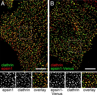
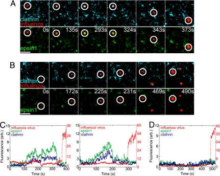
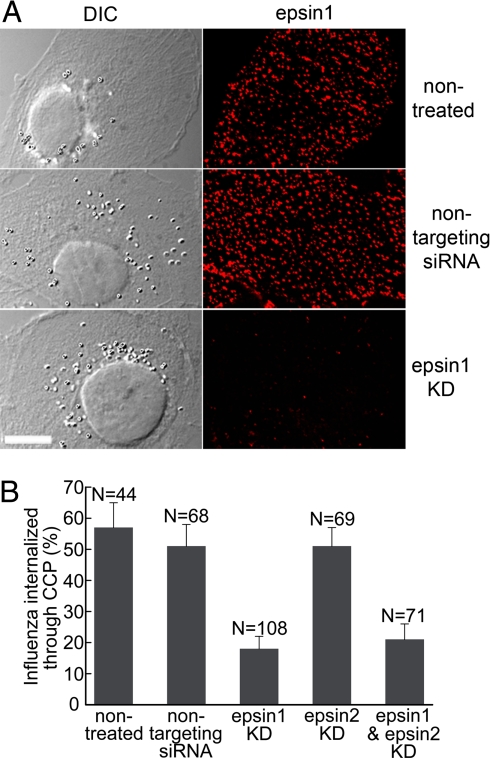
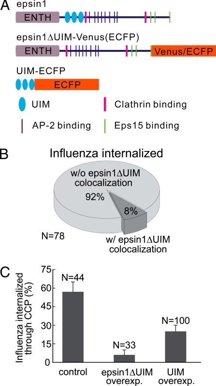
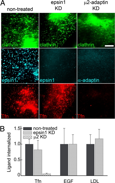
During clathrin-mediated endocytosis, adaptor proteins recognize specific internalization signals on cargo receptors, either recruiting cargos into clathrin-coated pits (CCPs) or initiating clathrin-coat assembly around the cargo molecules. Here, we identify epsin 1, a clathrin-, ubiquitin-, and phospholipid-interacting protein, as a cargo-specific adaptor for influenza virus entry through the clathrin-mediated pathway. Using live-cell imaging to monitor the entry of individual virus particles, we observed recruitment of epsin 1 to the binding sites of influenza viruses in synchrony with the assembly of CCPs. Epsin 1 knockdown by siRNA significantly inhibited the clathrin-mediated endocytosis of the influenza virus and caused the majority of the virus particles to enter through a clathrin-independent pathway. The same treatment did not affect the entry of several classical ligands for clathrin-mediated endocytosis, including transferrin, LDL, and EGF. Overexpression of the dominant-negative epsin 1 mutant lacking the ubiquitin-interaction motifs nearly completely blocked the clathrin-mediated entry of the influenza virus without affecting transferrin uptake. These results suggest that epsin 1 functions as a cargo-specific adaptor for the clathrin-mediated entry of the influenza virus.


Conflict of interest statement

The authors declare no conflict of interest.

Figures

Fig. 1.
Fig. 1.
Intracellular distribution of epsin 1. (A Upper) Immunofluorescence image of endogenous epsin 1 (red) and endogenous clathrin (green). (Lower) Magnified view of the boxed region. Shown are the epsin 1(Left), clathrin (Center), and overlaid (Right) images. (B Upper) Overlay of the Venus fluorescence image of epsin 1-Venus (green) and the immunofluorescence image of endogenous clathrin (red). The cell was transiently transfected with epsin 1-Venus. (Lower) Magnified view of the boxed region. Shown are the epsin 1-Venus (Left), clathrin (Center), and overlaid (Right) images. (Scale bars,10 μm.)
Fig. 2.
Fig. 2.
Influenza colocalized with epsin 1 during clathrin-mediated endocytosis. (A) Snapshots of a virus (red, circled) internalized through a CCP, colocalizing with both epsin 1 (green) and clathrin (cyan), and fused with an endosome. (Top) Clathrin and the virus channels. (Bottom) Epsin 1 and the virus channels of the three-color movie. Time indicates how long the virus was bound to the cell. (B) Snapshots of a virus (red, circled) that was internalized without colocalizing with either epsin 1 (green) or clathrin (cyan) and fused with an endosome. (Scale bars, 2.5 μm) (C) Fluorescence time traces of the DiD (red), epsin 1-Venus (green), and clathrin-ECFP (blue) signals associated with two virus particles internalized through clathrin-mediated endocytosis. (Left) Corresponds with the virus shown in A. (Right) Fluorescence time traces of a different virus, of which the snapshots are not shown. (D) Fluorescence time traces of the DiD, epsin 1-Venus, and clathrin-ECFP signals associated with a virus particle internalized through clathrin-independent mechanism, the snapshots of which are shown in B.
Fig. 3.
Fig. 3.
Epsin 1 knockdown inhibited the clathrin-mediated endocytosis of the influenza virus. (A) Efficient epsin 1 knockdown by siRNA. (Left) Differential interference contrast images of the cells. (Right) Corresponding immunofluorescence images of endogenous epsin 1. Shown are nontreated cells (Top), nontargeting siRNA-treated cells (Middle), and epsin 1 siRNA treated cells (Bottom). (Scale bar, 10 μm.) (B) The fraction of successfully internalized influenza virus particles that entered through clathrin-mediated endocytosis in nontreated, nontargeting siRNA treated, epsin 1 knockdown, epsin 2 knockdown, and epsin 1 and epsin 2 double knockdown cells.
Fig. 4.
Fig. 4.
Overexpression of epsin1ΔUIM or tandem UIM motifs inhibited the clathrin-mediated endocytosis of the influenza virus. (A) Schematic illustration of wild-type epsin 1, the epsin 1 mutant lacking the UIM motifs (epsin1ΔUIM) fused with Venus or ECFP, and the fragment that contains only the three tandem UIM motifs fused with ECFP. (B) The UIMs of epsin 1 is critical for its recruitment to the virus binding sites. In cells expressing epsin1ΔUIM-Venus, most of the virus particles did not show colocalization with epsin1ΔUIM-Venus before their internalization. (C) The fraction of influenza virus particles that entered through clathrin-mediated endocytosis in cells overexpressing epsin1ΔUIM or UIM. Here, cells were cotransfected with clathrin-EYFP and one of the two proteins: epsin1ΔUIM-ECFP or UIM-ECFP. Cells that showed overexpression of epsin1ΔUIM-ECFP or UIM-ECFP were chosen for analysis, and the results were compared with that observed in control cells not expressing epsin1ΔUIM-ECFP or UIM-ECFP.
Fig. 5.
Fig. 5.
Epsin 1 knockdown did not affect transferrin (TFn), EGF, or LDL uptake. (A) Fluorescence images of clathrin (Top), epsin 1 or AP-2 (Middle), and transferrin (Bottom) in nontreated (Left), epsin 1 knockdown (Center), and μ2-adaptin knockdown (Right) cells. The clathrin, epsin 1, and AP-2 signals were detected by immunofluorescence. To image internalized transferrin, cells were incubated with Alexa 633-labeled transferrin at 37°C for 15 min to allow internalization, and then cell-surface transferrin was removed by acid buffer wash. (Scale bar, 10 μm.) Similar assays were used for EGF and LDL (Fig. S8). (B) The amount of internalized transferrin, EGF, and LDL in epsin 1 or μ2-adaptin knockdown cells in comparison with that in control cells. The total fluorescence intensity of the ligands was normalized against that in control cells and used to quantify the internalization amount. Results were averaged over 10–15 cells in each case.

Similar articles

Cited by

References

    1. Conner SD, Schmid SL. Regulated portals of entry into the cell. Nature. 2003;422:37–44. - PubMed
    1. Bonifacino JS, Lippincott-Schwartz J. Coat proteins: Shaping membrane transport. Nat Rev Mol Cell Biol. 2003;4:409–414. - PubMed
    1. Traub LM. Sorting it out: AP-2 and alternate clathrin adaptors in endocytic cargo selection. J Cell Biol. 2003;163:203–208. - PMC - PubMed
    1. Robinson MS. Adaptable adaptors for coated vesicles. Trends Cell Biol. 2004;14:167–174. - PubMed
    1. Conner SD, Schmid SL. Differential requirements for AP-2 in clathrin-mediated endocytosis. J Cell Biol. 2003;162:773–779. - PMC - PubMed

Publication types

MeSH terms

LinkOut - more resources